Advantages:
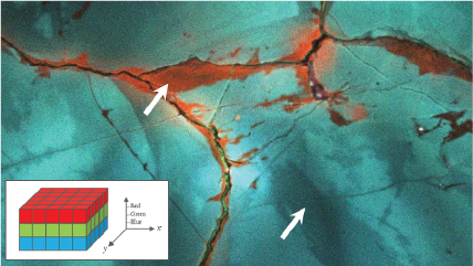
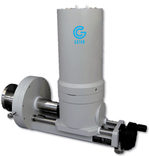
The ChromaCL2™ iBSED detector is a color cathodoluminescence imaging system for use in scanning electron microscopes (SEMs). The system is suitable for many petrographic and earth science applications where color cathodoluminescence (CL) imaging can reveal geochemical processes.
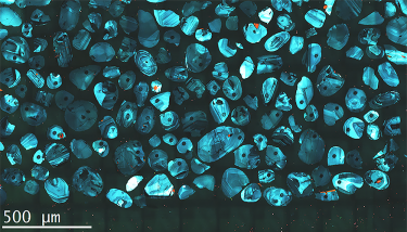
- Reveal macro- and micro-texture in sedimentary rocks including sediment source, degree of compaction, diagenetic history, differentiation of authigenic and detrital minerals, cementation history, and provenance
- Collect topographic, composition, and color CL images simultaneously in a single pass of the electron beam
- Simple operation
- Color cathodoluminescence images aid the interpretation of geochemistry changes
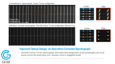
- High-efficiency detection enables fastest time-to-data, images with highest spatial resolution and reduces the likelihood of misinterpreting data as a result of e-beam damage
- Carbonate imaging kit to overcome phosphorescence from some carbonate minerals in scanned CL images
- Effectively unlimited field of view with field stitching software module


")