什么是系列连续切片成像?
系列连续切片扫描电子显微术(SBEM、SBSEM 和 SBFSEM)是从样品中重复获取高分辨率 3D 图像的一种方式。此方法尤擅在 X,Y,Z 坐标中进行纳米级分辨率的大视野成像。SBEM 通常依赖于扫描电子显微镜 (SEM) 中安装的原位超薄切片机。SEM 将收集切面图像,然后超薄切片机切割样品,暴露要成像的下一层,逐层切割,每层薄至 15 nm。
SBEM 的优势
功能 | 优势 |
---|---|
批量检验多个量级的结构 | 保留整个大数据集的细节层次 |
实现无中断的高产出 | 有助于加快样品分析和减少人为错误 |
展现所观察超微结构的特征 | 让您能够从一个样品中得到更全面的结果 |
控制切片和获取流程的各个方面 | 提供充分的灵活性,允许针对给定样品进行优化 |
消除截面损失、损坏和畸变 | 避免禁止的流程来修正损坏和畸变 |
用途
神经系统科学 | 细胞生物学 | 材料科学 | 其他 |
---|---|---|---|
|
|
|
|
神经系统科学
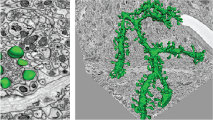
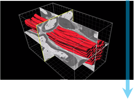
对 15,625 μm³ (25 x 25 x 25 μm) 体积数据集中的树突进行 3D 重建,该数据集包含 3View® 系统生成的 500 个小鼠小脑序列图像。树突结构(绿色)、终扣(黄色)和囊泡(红色)。插页图像,从左上方顺时针方向:树突共焦图像;渲染为体积模型的线框曲线;带突触的超高分辨率树突棘模型;显示线框曲线的图像。
细胞生物学:补充共焦
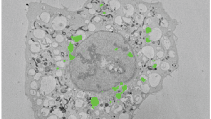
在无血清介质中饥饿 2 小时的 HeLa 细胞(能稳定表达刻蚀坐标玻璃底平皿盖玻片上生长的 LC-GFP)以及共焦显微镜识别的相关细胞的图像。然后,细胞经过原位处理以用于电子显微术,使用氢氟酸溶解盖玻片上的环氧树脂。再次在树脂坯块和 3View 系统生成的连续序列图像中识别细胞。
细胞生物学:发展
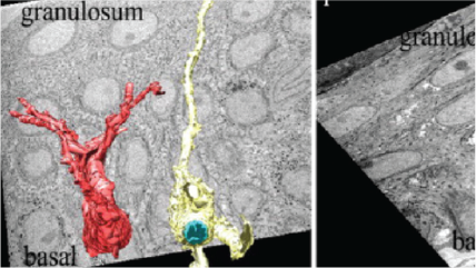
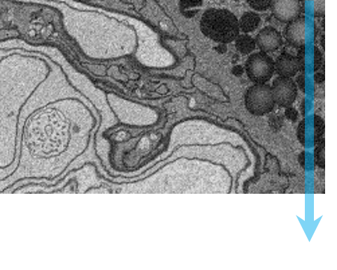
左上:高分辨率小鼠肾脏 8192 x 8192 像素图像,像素大小为 1.5 nm。上排中间:大视野小鼠肾脏 8192 x 8192 像素图像,像素大小为 80 nm。右:高压冷冻技术制备的秀丽隐杆线虫;高分辨率小鼠肾脏 4096 x 4096 像素图像,像素大小为 25 nm。样品由加州大学伯克利分校的 Kent McDonald 提供。左下:小鼠坐骨神经 2048 x 2048 像素图像,像素大小为 5 nm。下排中间:小鼠坐骨神经轴突 3D 可视化图像。
材料科学
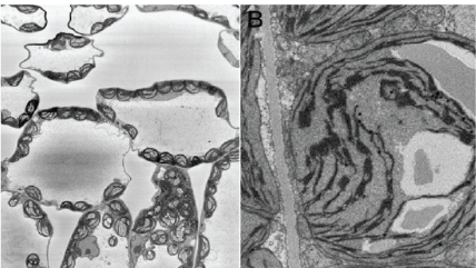
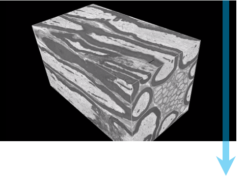
左上和左下:3View 系统生成的铝表面阳极处理层图像。中间:3View 系统生成的带有锰颗粒的铝合金 3D 可视化图像。3D 数据集包含像素大小为 15 nm 且切割厚度为 15 nm 的 1,000 个 1024 x 1024 序列图像。使用 3D 可视化插件在 DigitalMicrograph® 中创建的 3D 模型。
系列连续切片成像的工作流程
|
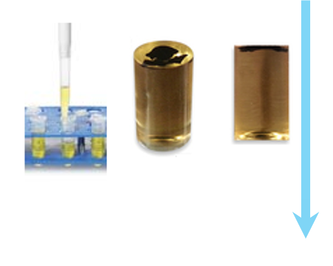
第 1 步:样品制备 固定典型生物样品,使用造影剂对样品进行着色,将样品嵌入树脂中使其保持稳定。 |
![]() |
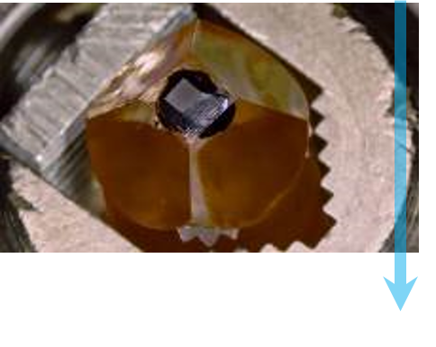
第 2 步:安装并转移到 SEM 中 将样品修整为需要的大小,固定在铝销上,可通过溅射镀膜方法镀上一层金薄膜。将样品销放在 3View 系统中,与金刚石刀接触,门关闭并收回。 |
![]() |
第 3 步:优化成像 用户选择合适的电子束条件,类似于标准 SEM 所用的条件,但同时应考虑 Z 方向上的效应。选择合适的放大倍数、像素数和停留时间,以达到理想的图像分辨率、视野和获取时间。 |
![]() |
第 4 步:自动成像 用户可自行选择电子束条件来获取序列图像。生成每个图像之前,切割刀都会剥离一层样品表面。 |
![]() |
第 5 步:分析 图像堆叠形成 3D 数据集,可使用 DigitalMicrograph® 或第三方软件处理和查看。此时即可进行分段和定量。 |
DigitalMicrograph,或称为 Gatan Microscopy Suite,驱动您的电子相机和其他附件以支持一系列重要应用,包括断层扫描、原位、谱学和衍射成像等。
3D-CLEM reveals that a major portion of mitotic chromosomes is not chromatin
Booth, D. G.; Beckett, A. J.; Molina, O.; Samejima, I.; Masumoto, H.; Kouprina, N.; Larionov, V.; Prior, I. A.; Earnshaw, W. C.
Holcomb, P. S.; Hoffpauir, B. K.; Hoyson, M. C.; Jackson, D. R.; Deerinck, T. J.; Marrs, G. S.; Dehoff, M.; Wu, J.; Ellisman, M. H.; Spirou, G. A.
High-throughput 3D visualization of large volumes at high resolution by 3View
Dohnalkova, A.; Kennedy, D.; Mancuso, J.; Marshall, M.; Mainwaring, P.; Fredrickson, J.
Lead asparate, an en bloc contrast stain particularly useful for ultrastructural enzymology
Walton, J.
Morphology of phagophore precursors by correlative light-electron microscopy
Gudmundsson, S. R.; Kallio, K. A.; Vihinen, H.; Jokitalo, E.; Ktistakis, N.; Eskelinen, E. L.
Tanaka, T.; Ohno, N.; Osanai, Y.; Saitoh, S.; Thai, T. Q.; Nishimura, K.; Shinjo, T.; Takemura, S.; Tatsumi, K.; Wanaka, A.
Mitochondrial morphology and function: two for the price of one!
Faitg, J.; Davey, T.; Turnbull, D. M.; White, K.; Vincent, A. E.
Ultrastructural variability of the juxtacanalicular tissue along the inner wall of Schlemm’s canal
Koudouna, E.; Young, R. D.; Overby, D. R.; Ueno, M.; Kinoshita, S.; Knupp, C.; Quantock, A. J.
Hashimoto, T.; Thompson, G. E.; Zhou, X.; Withers, P. J.
The biosynthesis of infrared-emitting quantum dots in allium fistulosum
Green, M.; Haigh, S. J.; Lewis, E. A.; Sandiford, L.; Burkitt-Gray, M.; Fleck, R.; Vizcay-Barrena, G.; Jensen, L.; Mirzai, H.; Curry, R. J.; Dailey, L. -A.
Borrett, S.; Hughes, L.
Kreshuk, A.; Walecki, R.; Koethe, U.; Gierthmuehlen, M.; Plachta, D.; Genoud, C.; Haastert-Talini, K.; Hamprecht, F. A.
Froud, K. E.; Wong, A. C. Y.; Cederholm, J. M. E.; Klugmann, M.; Sandow, S. L.; Julien, J. -P.; Ryan, A. F.; Housley, G. D.
Challenges of microtome-based serial block-face scanning electron microscopy in neuroscience
Wanner, A. A.; Kirschmann, M. A.; Genoud, C.
High-resolution whole-brain staining for electron microscopic circuit reconstruction
Mikula, S.; Denk, W.
Comparison and combination of imaging techniques for three dimensional analysis of electrical trees
Schurch, R.; Rowland, S.M.; Bradley, R.S.; Withers, P.J.
The Lowe syndrome protein OCRL1 is required for endocytosis in the zebrafish pronephric tubule
Oltrabella, F.; Pietka, G.; Ramirez, I. B.-R.; Mironov, A.; Starborg, T.; Drummond, I. A.; Hinchliffe, K. A.; Lowe, M.
Starborg, T.; Kadler, K. E.
Hondow, N.; Brown, M. R.; Starborg, T.; Monteith, A. G.; Brydson, R.; Summers, H. D.; Rees, P.; Brown, A.
Three-dimensional architecture of podocytes revealed by block-face scanning electron microscopy
Ichimura, K.; Miyazaki, N.; Sadayama, S.; Murata, K.; Koike, M.; Nakamura, K. -I.; Ohta, K.; Sakai, T.
Analysis of primary cilia in the developing mouse brain
Paridaen, J. T. M. L.; Huttner, W. B.; Wilsch-Bräuninger, M.
How to bury the dead: elimination of apoptotic hair cells from the hearing organ of the mouse
Anttonen, T.; Belevich, I.; Kirjavainen, A.; Laos, M.; Brakebusch, C.; Jokitalo, E.; Pirvola, U.
Developing 3D SEM in a broad biological context
Kremer, A.; Lippens, S.; Bartunkova, S.; Asselbergh, B.; Blanpain, C.; Fendrych, M.; Goossens, A.; Holt, M.; Janessens, S.; Krols, M.; Larsimont, J. -C.; Mc Guire, C.; Nowack, M. K.; Saelens, X.; Schertel, A.; Schepens, B.; Slezak, M.; Timmerman, V.; Theunis, C.; Van Brempt, R.; Visser, Y.; Guerin, C. J.
Application of FEA to image-based models of electrical trees with uniform conductivity
Rowland, S. M.; Schurch, R.; Pattouras, M.; Li, Q.
RBM3 mediates structural plasticity and protective effects of cooling in neurodegeneration
Peretti, D.; Bastide, A.; Radford, H.; Verity, N.; Molloy, C.; Martin, M. G.; Moreno, J. A.; Steinert, J. R.; Smith, T.; Dinsdale, D.; Willis, A. E.; Mallucci, G. R.
Quantitative analysis of mouse pancreatic islet architecture by serial block-face SEM
Pfeifer, C. R.; Shomorony, A.; Aronova, M. A.; Zhang, G.; Cai, T.; Xu, H.; Notkins, A. L.; Leapman, R. D.
Advanced imaging and tissue engineering of the human limbal epithelial stem cell niche
Massie, I.; Dziasko, M.; Kureshi, A.; Levis, H. J.; Morgan, L.; Neale, M.; Sheth, R.; Tovell, V. E.; Vernon, A. J.; Funderburgh, J. L.; Daniels, J. T.
Kempen, P. J.; Kircher, M. F.; de la Zerda, A.; Zavaleta, C. L.; Jokerst, J. V.; Mellinghoff, I. K.; Gambhir, S. S.; Sinclair, R.
Pinali, C.; Holt, C. M.; Bennett, H. J.; Davenport, J. B.; Walker, R.; Murfitt, L.; Allan, L. J.; Malik, N.; Kitmitto, A.
Automated analysis of spine dynamics on live CA1 pyramidal cells
Blumer, C.; Vivien, C.; Genoud, C.; Perez-Alvarez, A.; Wiegert, J. S.; Vetter, T.; Oertner, T. G.
Automated analysis of spine dynamics on live CA1 pyramidal cells
Blumer, C.; Vivien, C.; Genoud, G.; Perez-Alvarez, A.; Wiegert, J. S.; Vetter, T.; Oertner, T. G.
Ju, W. K.; Kim, K. Y.; Noh, Y. H.; Hoshijima, M.; Lukas, T. J.; Ellisman, M. H.; Weinreb, R. N.; Perkins, G. A.
Finite element analysis of the pressure-induced deformation of Schlemm’s canal endothelial cells
Vargas-Pinto, R.; Lai, J.; Gong, H.; Ethier, C. R.; Johnson, M.
Serial block face-scanning electron microscopy: a method to study retinal degenerative phenotypes
Mustafi, D.; Kikano, S.; Palczewski, K.
Serial block face-scanning electron microscopy: A method to study retinal degenerative phenotypes
Mustafi, D.; Kikano, S.; Palczewski, K.
Staining and embedding of human chromosomes for 3-d serial block-face scanning electron microscopy
Yusuf, M.; Chen, B.; Hashimoto, T.; Estandarte, A. K.; Thompson, G.; Robinson, I.
Three-dimensional reconstruction of skeletal muscle extracellular matrix ultrastructure
Gillies, A. R.; Bushong, E. A.; Deerinck, T. J.; Ellisman, M. H.; Lieber, R. L.
The versatile electron microscope: An ultrastructural overview of autophagy
Biazik, J.; Vihinen, H.; Anwar, T.; Jokitalo, E.; Eskelinen, E.
Mission (im)possible – mapping the brain becomes a reality
Eberle, A. L.; Selchow, O.; Thaler, M.; Zeidler, D.; Kirmse, R.
Dynein light intermediate chains maintain spindle bipolarity by functioning in centriole cohesion
Jones, L. A.; Villemant, C.; Starborg, T.; Salter, A.; Goddard, G.; Ruane, P.; Woodman, P. G.; Papalopulu, N.; Woolner, S.; Allan, V. J.
Urwyler, O.; Izadifar, A.; Dascenco, D.; Petrovic, M.; He, H.; Ayaz, D.; Kremer, A.; Lippens, S.; Baatsen, P.; Guérin, C. J.; Schmucker, D.
Pinali, C.; Kitmitto, A.
En bloc staining with hydroquinone treatment for block face imaging
Togo, A.; Ohta, K.; Higashi, R.; Nakamura, K.
Three dimensional imaging of electrical trees in micro and nano-filled epoxy resin
Schurch, R.; Rowland, S. M.; Bradley, R. S.; Hashimoto, T.; Thompson, G. E.; Withers, P. J.
Three dimensional imaging of electrical trees in micro and nano-filled epoxy resin
Schurch, R.; Rowland, S.M.; Bradley, R.S.; Hashimoto, T.; Thompson, G.E.; Withers, P.J.
Finite-size effects in the 3D reconstruction and morphological analysis of porous polymers
Müllner, T.; Zankel, A.; Svec, F.; Tallarek, U.
Salloum, R. H.; Chen, G.; Velet, L.; Manzoor, N. F.; Elkin, R.; Kidd, GJ.; Coughlin, J.; Yurosko, C.; Bou-Anak, S.; Azadi, S.; Gohlsch, S.; Schneider, H.; Kaltenbach, J.A.
Salloum, R. H.; Chen, G.; Velet, L.; Manzoor, N. F.; Elkin, R.; Kidd, G. J.; Coughlin, J.; Yurosko, C.; Bou-Anak, S.; Azadi, S.; Gohlsch, S.; Schneider, H.; Kaltenbach, J.A.
A quantitative three-dimensional in situ study of a short fatigue crack in a magnesium alloy
Marrow, T. J.; Mostafavi, M.; Hashimoto, T.; Thompson, G. E.
A quantitative three-dimensional in situ study of a short fatigue crack in a magnesium alloy
Marrow, T. J.; Mostafavi, M.; Hashimoto, T.; Thompson, G. E.
Rhinomonas nottbecki n. sp. (cryptomonadales) and molecular phylogeny of the family pyrenomonadaceae
Majaneva, M.; Remonen, I.; Rintala, J. M.; Belevich, I.; Kremp, A.; Setälä, O.; Jokitalo, E.; Blomster, J.
Arabidopsis NAC45/86 direct sieve element morphogenesis culminating in enucleation
Furuta, K. M.; Yadav, S. R.; Lehesranta, S.; Belevich, I.; Miyashima, S.; Heo, J.; Vatén, A.; Lindgren, O.; De Rybel, B.; Van Isterdael, G.; Somervuo, P.; Lichtenberger, R.; Rocha, R.; Thitamadee, S.; Tähtiharju, S.; Auvinen, P.; Beeckman, T.; Jokitalo, E.; Helariutta, Y.
Tsai, W. T.; Hassan, A.; Sarkar, P.; Correa, J.; Metlagel, Z.; Jorgens, D. M.; Auer, M.
Busskamp, V.; Krol, J.; Nelidova, D.; Daum, J.; Szikra, T.; Tsuda, B.; Jüttner, J.; Farrow, K.; Scherf, B. G.; Alvarez, C. P.; Genoud, C.; Sothilingam, V.; Tanimoto, N.; Stadler, M.; Seeliger, M.; Stoffel, M.; Filipowicz, W.; Roska, B.
Miyazaki, N.; Esaki, M.; Ogura, T.; Murata, K.
Heras-Bautista, C. O.; Katsen-Globa, A.; Schloerer, N. E.; Dieluweit, S.; El Aziz OM, A.; Peinkofer, G.; Attia, W. A.; Khalil, M.; Brockmeier, K.; Hescheler, J.; Pfannkuche, K.
Miyazaki, N.; Esaki, M.; Ogura, T.; Murata, K.
Differential roles of microglia and monocytes in the inflamed central nervous system
Yamasaki, R.; Lu, H.; Butovsky, O.; Ohno, N.; Rietsch, A.M.; Cialic, R.; Wu, P. M.; Doykan, C. E.; Lin, J.; Cotleur, A. C.; Kidd, G.; Zorlu, M. M.; Sun, N.; Hu, W.; Liu, L.; Lee, J. C.; Taylor, S. E.; Uehlein, L.; Dixon, D.; Gu, J.; Floruta, C. M.; Zhu, M.; Charo, I. F.; Weiner, H. L.; Ransohoff, R. M.
Mitochondrial immobilization mediated by syntaphilin facilitates survival of demyelinated axons
Ohno, N.; Chiang, H.; Mahad, D. J.; Kidd, G. J.; Liu, L.; Ransohoff, R. M.; Sheng, Z. H.; Komuro, H. Trapp, B. D.
Cooperrider, J.; Furmaga, H.; Plow, E.; Park, H. J.; Chen, Z.; Kidd, G.; Baker, K. B.; Gale, J. T.; Machado, A. G.
Hughes, A. E.; Trinchi, A.; Chen, F. F.; Yang, Y. S.; Cole, I. S.; Sellaiyan, S.; Carr, J.; Lee, P. D.; Thompson, G. E.; Xiao, T. Q.
Serial sectioning methods for 3D investigations in materials science
Zankel, A.; Wagner, J.; Poelt, P.
Lipke, E.; Hörnschemeyer, T.; Pakzad, A.; Booth, C. R.; Michalik, P.
Chen, B.; Hashimoto, T.; Vergeer, F.; Burgess, A.; Thompson, G.; Robinson, I.
Pinali, C.; Bennett, H. J.;Davenport, J. B.; Caldwell, J. L.; Trafford, A. W.; Kitmitto, A.
Lipke, E.; Hörnschemeyer, T.; Pakzad, A.; Booth, C. R.; Michalik, P.
Pinali, C.; Bennett, H. J.; Davenport, J. B.; Caldwell, J. L.; Trafford, A. W.; Kitmitto, A.
Transcellular degradation of axonal mitochondria
Davis, C. O.; Kim, K.; Bushong, E. A.; Mills, E. A.; Boassa, D.; Shih, T.; Kinebuchi, M.; Phan, S.; Zhou, Y.; Bihlmeyer, N. A.; Nguyen, J. V.; Jin, Y.; Ellisman, M. H.; Marsh-Armstrong, N.
Revelation of intertwining organic and inorganic fractal structures in polymer coatings
Hughes, A. E.; Trinchi, A.; Chen, F. F.; Yang, Y. S.; Cole, I. S.; Sellaiyan, S.; Carr, J.; Lee, P. D.; Thompson, G. E.; Xiao, T. Q.
Dziasko, M. A.; Armer, H. E.; Levis, H. J.; Shortt, A. J.; Tuft, S.; Daniels, J. T.
Wu, J.; Bakerink, K. J.; Evangelista, M. E.; Thomas, G. H.
ER sheet persistence is coupled to myosin 1c–regulated dynamic actin filament arrays
Joensuu, M.; Belevich, I.; Rämö, O.; Nevzorov, I.; Vihinen, H.; Puhka, M.; Witkos, T. M.; Lowe, M.; Vartiainen, M. K.; Jokitalo, E.
Gillies, A.; Lieber, R.
Serial block face scanning electron microscopy-the future of cell ultrastructure imaging
Hughes, L.; Hawes, C.; Monteith, S.; Vaughan, S.
An enteroendocrine cell-enteric glia connection revealed by 3D electron microscopy
Bohórquez, D. V.; Samsa, L. A.; Roholt, A.; Medicetty, S.; Chandra, R.;Liddle, R. A.
Pingel, J.; Lu, Y.; Starborg, T.; Fredberg, U.; Langberg, H.; Nedergaard, A.; Weis, M.; Eyre, D.; Kjaer, M.; Kadler, K. E.
Resolution of the three dimensional structure of components of the glomerular filtration barrier
Arkill, K. P; Qvortrup, K.; Starborg, T.; Mantell, J. M.; Knupp, C.; Michel, C. C.; Harper, S. J.; Salmon, A. H. J.; Squire, J. M.; Bates, D. O.; Neal, C. R.
Three-dimensional aspects of matrix assembly by cells in the developing cornea
Young, R. D.; Knupp, C.; Pinali, C.; Png, K. M. Y.; Ralphs, J. R.; Bushby, A. J.; Starborg,T.; Kadler, K. E.; Quantock, A. J.
Wilke, S. A.; Raam, T.; Antonios, J. K.; Bushong, E. A.; Koo, E. H.; Ellisman, M. H.; Ghosh, A.
Resolution of three dimensional structure of components of the glomerular filtration barrier
Arkill, K. P.; Qvortrup, K.; Starborg, T.; Mantell, J. M.; Knupp, C.; Michel, C. C.; Harper, S. J.; Salmon, A. H. J.; Squire, J. M.; Bates, D. O.; Neal, C. R.
Exploring the third dimension: Volume electron microscopy comes of age
Peddie, C. J.; Collinson, L. M.
Hughes, L.; Towers, K.; Starborg, T.; Gull, K.; Vaughan, S.
The protein quality control system manages plant defence compound synthesis
Pollier, J.; Moses, T.; González-Guzmán, M.; De Geyter, N.; Lippens, S.; Bossche, R. V.; Marhavý, P.; Kremer, A.; Morreel, K.; Guérin, C. J.; Tava, A.; Oleszek, W.; Thevelein, J. M.; Campos, N.; Goormachtig, S.; Goossens, A.
Nonmuscle myosin II powered transport of newly formed collagen fibrils at the plasma membrane
Kalson, N. S.; Starborg, T.; Lu, Y.; Mironov, A.; Humphries, S. M.; Holmes, D. F.; Kadler, K. E.
Widespread mitochondrial depletion via mitiphagy does not compromise necroptosis
Tait, S. W.; Oberst, A.; Quarato, G.; Milasta, S.; Haller, M.; Wang, R.; Karvela, M.; Ichim, G.; Yatim, N.; Albert, M. L.; Kidd, G.; Wakefield, R.; Frase, S.; Krautwald, S.; Linkermann, A.; Green, D. R.
Pinali, C.; Bennett, H.; Davenport, J. B.; Trafford, A. W.; Kitmitto, A.
Kuwajima, M.; Spacek, J.; Harris, K. M.
Trueman, A.; Knight, S.; Colwell, J.; Hashimoto, T.; Carr, J.; Skeldon, P.; Thompson, G.
Connectomic reconstruction of the inner plexiform layer in the mouse retina
Helmstaedter, M.; Briggman, K. L.; Turaga, S. C.; Jain, V.; Seung, H. S.; Denk, W.
Dwyer, L.; Robson, J.; da Fonseca, J. Q.; Kamp, N.; Hashimoto, T.; Thompson, G. E.
Construction of a polarized neuron
Holcomb, P. S.; Deerinck, T. J.; Ellisman, M. H.; Spirou, G. A.
Hashimoto, T.; Thompson, G. E.; Curioni, M.; Zhou, X. R.; Skeldon, P.
Three dimensional electron microscopy of cellular organelles by serial block face SEM and ET
Vihinen, H.; Belevich, I.; Jokitalo, E.
Wong, J.; Baddeley, D.; Bushong, E. A.; Yu, Z.; Ellisman, M. H.; Hoshijima, M.; Soeller, C.
Functional development of the olfactory system in zebrafish
Miyasaka, N.; Wanner, A. A.; Li, J.; Mack-Bucher, J.; Genoud, C.; Yoshihara, Y.; Friedrich, R. W.
BMP signaling specifies the development of a large and fast CNS synapse
Xiao, L.; Michalski, N.; Kronander, E.; Gjoni, E.; Genoud, C.; Knott, G.; Schneggenburger, R.
DP2: Distributed 3D image segmentation using micro-labor workforce
Giuly, R. J.; Kim, K. Y.; Ellisman, M. H.
Shigetomi, E.; Bushong, E. A.; Haustein, M. D.; Tong, X.; Jackson-Weaver, O.; Kracun, S.; Xu, J.; Sofroniew, M. V.; Ellisman, M. H.; Khakh, B. S.
Analyzing the structure and function of neuronal circuits in zebrafish
Friedrich, R. W.; Genoud, C.; Wanner, A. A.
WDR81 is necessary for purkinje and photoreceptor cell survival
Traka, M.; Millen, K. J.; Collins, D.; Elbaz, B.; Kidd, G. J.; Gomez, C. M.; Popko, B.
Large-scale automatic reconstruction of neuronal processes from electron microscopy images
Kaynig, V.; Vazquez-Reina, A.; Knowles-Barley, S.; Roberts, M.; Jones, T. R.; Kasthuri, N.; Miller, E.; Lichtman, J.; Pfister, H.
Boassa, D.; Berlanga, M. L.; Yang, M. A.; Terada, M.; Hu, J.; Bushong, E. A.; Hwang, M.; Masliah, E.; George, J. M.; Ellisman, M. H.
Revealing the three dimensional internal structure of aluminium alloys
Thompson, G. E.; Hashimoto, T.; Zhong, X. L.; Curioni, M.; Zhou, X.; Skeldon, P.; Withers, P. J.; Carr, J. A.; Monteith, A. G.
Jurrus, E.; Watanabe, S.; Giuly, R. J.; Paiva, A. R.; Ellisman, M. H.; Jorgensen, E. M.; Tasdizen, T.
Wilke, S. A.; Antonios, J. K.; Bushong, E. A.; Badkoobehi, A.; Malek, E.; Hwang, M.; Terada, M.; Ellisman, M. H.; Ghosh, A.
Autosomal recessive retinitis pigmentosa E150K opsin mice exhibit photoreceptor disorganization
Zhang, N.; Kolesnikov, A. V.; Jastrzebska, B.; Mustafi, D.; Sawada, O.; Maeda, T.; Genoud, C.; Engel, A.; Kefalov, V. J.; Palczewski, K.
Starborg, T.; Kalson, N. S.; Lu, Y.; Mironov, A.; Cootes, T. F.; Holmes, D. F.; Kadler, K. E.
Paridaen, J. T. M. L.; Wilsch-Bräuninger, M.; Huttneremail, W. B.
Three-dimensional structure analysis and percolation properties of a barrier marine coating
Chen, B.; Guizar-Sicairos,M.; Xiong,G.; Shemilt, L.; Diaz, A.; Nutter, J.; Burdet,N.; Huo,S.; Mancuso, J.; Monteith,A.; Vergeer,F.; Burgess,A.; Robinson, I.
Anttonen, T.; Kirjavainen, A.; Belevich, I.; Laos, M.; Richardson, W. D.; Jokitalo, E.; Brakebusch, C.; Pirvola, U.
Müllner, T.; Zankel, A.; Mayrhofer, C.; Reingruber, H.; Höltzel, A.; Lv, Y.; Svec, F.; Tallarek, U.
Investigation of dealloying by ultra-high-resolution nanotomography
Hashimoto, T.; Curioni, M.; Zhou, X.; Mancuso, J.; Skeldon, P.; Thompson, G. E.
Schwartz, C. L.; Heumann, J. M.; Dawson, S. C.; Hoenger,A.
Prdm3 and Prdm16 are H3K9me1 methyltransferases required for mammalian heterochromatin integrity
Pinheiro, I.; Margueron, R.; Shukeir, N.; Eisold, M.; Fritzsch, C.; Richter, F. M.; Mittler, G.; Genoud, C.; Goyama, S.; Kurokawa, M.; Son, J.; Reinberg, D.; Lachner, M.; Jenuwein, T.
Puhka, M.; Joensuu, M.; Vihinen, H.; Belevich, I.; Jokitalo, E.
Zhuravleva, E.; Gut, H.; Hynx, D.; Marcellin, D.; Bleck, C. K.; Genoud, C.; Cron, P.; Keusch, J. J.; Dummler, B.; Esposti, M. D.; Hemmings, B. A.
Azeloglu, E.; Stothers, M.; Deerinck, T. J.; Falkenberg, C.; Chen, Y.; He, J. C.; Hone, J. C.; Leslie, L. M.; Ellisman, M. H.; Iyengar, R.
Extrusion of misfolded and aggregated proteins--a protective strategy of aging neurons?
Doehner, J.; Genoud, C.; Imhof, C.; Krstic, D.; Knuesel, I.
3D segmentation of SBFSEM images of neuropil by a graphic model over supervoxel boundaries
Andres, B.; Koethe, U.; Kroeger, T.; Helmstaedter, M.; Briggman, K. L.; Denk, W.; Hamprecht, F. A.
Differential modulation of retinal degeneration by Ccl2 and Cx3cr1 chemokine signalling
Luhmann, U. F.; Lange, C. A.; Robbie, S.; Munro, P. M.; Cowing, J. A.; Armer, H. E.; Luong, V.; Carvalho, L. S.; MacLaren, R. E.; Fitzke, F. W.; Bainbridge, J.W.; Ali, R. R.
Beam deceleration for block-face scanning electron microscopy of embedded biological tissue
Ohta, K.; Sadayama, S.; Togo, A.; Higashi, R.; Tanoue, R.; Nakamura, K.
Multivesicular exocytosis in rat pancreatic beta cells
Hoppa, M. B.; Jones, E.; Karanauskaite, J.; Ramracheya, R.; Braun, M.; Collins, S. C.; Zhang, Q.; Clark, A.; Eliasson, L.; Genoud, C.; Macdonald, P. E.; Monteith, A. G.; Barg, S.; Galvanovskis, J.; Rorsman, P.
Structural neurobiology: Missing link to a mechanistic understanding of neural computation
Denk, W.; Briggman, K. L.; Helmstaedter, M.
Giuly, R. J.; Martone, M. E.; Ellisman, M. H.
Volume electron microscopy for neuronal circuit reconstruction
Briggman, K. L.; Bock, D. D.
Yan, J.; Walker, C. G.; O'sullivan, M. J.; Bushong, E. A.; Ellisman, M. H.; Hoshijima, M.; Rajagopal, V.
High contrast en bloc staining of neuronal tissue for field emission scanning electron microscopy
Tapia, J. C.; Kasthuri, N.; Hayworth, K. J.; Schalek, R.; Lichtman, J. W.; Smith, S. J.; Buchanan, J.
Modern electron microscopy methods for C. elegans
Hall, D. H.; Hartwieg, E.; Nguyen, K. C.
Ou, H. D.; Kwiatkowski, W.; Deerinck, T. J.; Noske, A.; Blain, K. Y.; Land, H. S.; Soria, C.; Powers, C. J.; May, A. P.; Shu, X.; Tsien, R. Y.; Fitzpatrick, J. A. J.; Long, J. A.; Ellisman, M. H.; Choe, S.; O'Sheaemail, C. C.
Mustafi, D.; Kevany, B. M.; Genoud, C.; Okano, K.; Cideciyan, A. V.; Sumaroka, A.; Roman, A. J.; Jacobson, S. G.; Engel, A.; Adams, M. D.; Palczewski, K.
Motskin, M.; Müller, K. H.; Genoud, C.; Monteith, A. G.; Skepper, J. N.
Kalson, N. S.; Holmes, D. F.; Herchenhan, A.; Lu, Y.; Starborg, T.; Kadler, K. E.
Wong, J. I. S.; Baddeley, D.; Bushong, E. A.; Ellisman, M.; Hoshijima, M.; Soeller, C.
Fast extraction of neuron morphologies from large-scale SBFSEM image stacks
Lang, S.; Drouvelis, P.; Tafaj, E.; Bastian, P.; Sakmann, B.
Mun, J. Y.; Jeong, S. Y.; Kim, J. H.; Han, S. S.; Kim, I. H.
Basement membrane changes in capillaries of the ageing human retina
Powner, M. B.; Scott, A.; Zhu, M.; Munro, P. M.; Foss, A. J.; Hageman, G. S.; Gillies, M. C.; Fruttiger, M.
Cadherin-9 regulates synapse-specific differentiation in the developing hippocampus
Williams, M. E.; Wilke, S. A.; Daggett, A.; Davis, E.; Otto, S.; Ravi, D.; Ripley, B.; Bushong, E. A.; Ellisman, M. H.; Klein, G.; Ghosh, A.
High-accuracy neurite reconstruction for high-throughput neuroanatomy
Helmstaedter, M.; Briggman, K. L.; Denk, W.
Role of elastin anisotropy in structural strain energy functions of arterial tissue
Rezakhaniha, R.; Fonck, E.; Genoud, C.; Stergiopulos, N.
Modeling of flow in a polymeric chromatographic monolith
Koku, H.; Maier, R. S.; Czymmek, K. J.; Schure, M. R.; Lenhoff, A. M.
Quantitative Characterisation of microfiltration membranes by 3D reconstruction
Reingruber, H.; Zankel, A.; Mayrhofer, C.; Poelt, P.
Shu, X.; Lev-Ram, V.; Deerinck, T. J.; Qi, T.; Ramko, E. B.; Davidson, M. W.; Jin, Y. Ellisman, M. H.; Tsien, R. Y.
Wiring specificity in the direction-selectivity circuit of the retina
Briggman, K. L.; Helmstaedter, M.; Denk, W.
Nguyen, J. V.; Soto, I.; Kim, K.; Bushong, E. A.; Oglesby, E.; Valiente-Soriano, F. J.; Yang, Z.; Davis, C. O.; Bedont, J. L.; Son, J. L.; Wei, J. O.; Buchman, V. L.; Zack, D. J.; Vidal-Sanz, M.; Ellisman, M. H.; Marsh-Armstrong, N.
Environmental scanning electron microscopy (ESEM)--a versatile tool in studying plants.
Stabentheiner, E.; Zankel A.; Pölt P.
Jurrus, E.; Paiva, A. R.; Watanabe, S.; Anderson, J. R.; Jones, B. W.; Whitaker, R. T.; Jorgensen, E. M.; Marc, R. E.; Tasdizen, T.
Reconstruction of a neuron from SBFSEM: tools, reliability, accuracy and efficiency
Drouvelis, P.; Kurz, T.; Bastian, P.; Sakmann, B.; Lang, S.
Deerinck, T. J.; Bushong, E. A.; Lev-Ram, V.; Shu, X.; Tsien, R. Y.; Ellisman, M. H.
Structure-function studies of blood and air capillaries in chicken lung using 3D electron microscopy
West, J. B.; Fu, Z.; Deerinck, T. J.; Mackey, M. R.; Obayashi, J. T.; Ellisman, M.H.
Imaging transient blood vessel fusion events in zebrafish by correlative volume electron microscopy
Armer, H. E. J.; Mariggi, G.; Png, K. M. Y.; Genoud, C.; Monteith, A. G.; Bushby, A. J.; Gerhardt, H.; Collinson, L. M.
Saetzler, K.; McCanny, P.; Rodriguez, E.P.; Horstmann, H.; Bruno, R.M.; Denk, W.
Axon tracking in serial block-face scanning electron microscopy
Jurrus, E.; Hardy, M.; Tasdizen, T.; Fletcher, P. T.; Koshevoy, P.; Chien, C. B.; Denk, W.; Whitaker, R.
Mishchenko, Y.
Three-dimensional reconstruction methods for Caenorhabditis elegans ultrastructure
Müller-Reichert, T.; Mancuso, J.; Lich, B.; McDonald, K.
Ultramicrotomy in the ESEM, a versatile method for materials and life sciences
Zankel, A.; Kraus, B.; Poelt, P.; Schaffer, M.; Ingolic, E.
Genoud, C.; Mancuso, J.; Monteith, S.; Kraus, B.
Contour-propagation algorithms for semi-automated reconstruction of neural processes
Macke, J. H.; Maack, N.; Gupta, R.; Denk, W.; Schölkopf, B.; Borst, A.
Segmentation of SBFSEM volume data of neural tissue by hierarchical classification
Andres, B.; Köthe, U.; Helmstaedter, M.; Denk, W.; Hamprecht, F. A.
Alpha-herpesvirus infection induces the formation of nuclear actin filaments
Feierbach, B.; Piccinotti, S.; Bisher, M.; Denk, W.; Enquist, L. W.
Serial block-face scanning electron microscopy to reconstruct three-dimensional tissue nanostructure
Denk, W.; Horstmann, H.