シリアルブロックフェイスイメージングとは
シリアルブロックフェイス走査電子顕微鏡法(SBEM、SBSEM、およびSBFSEM)は、試料から高解像度の三次元画像を再現可能な方法で取得する手法です。この手法は、ナノメートルレベルの分解能でX,Y,Zの広視野のイメージングを行う場合に特に適しています。通常、SBEMは、走査型電子顕微鏡(SEM)内部に組み込まれたin-situウルトラミクロトームを使用します。SEMが断面画像を収集すると、ウルトラミクロトームが15 nmという薄さで試料を切り出して、像観察を行う次の層を露出させます。
SBEMのメリット
機能 | メリット |
---|---|
複数桁のスケールにわたり構造を観察 | 大きなデータセット全体にわたり詳細なデータを取得 |
操作不要で高スループットを実現 | 試料分析を高速化し、人為的なミスを軽減 |
観察された超微細構造の厚さ方向の関係を解明 | 1つの試料からより包括的な結果を取得 |
セクショニングと像取得のあらゆるプロセスを管理 | 試料に対して条件を最適化するための完全な柔軟性を提供 |
切片の損失、損傷、変形の可能性を排除 | 損傷や変形を補正するために必要な処理を回避 |
用途
神経科学 | 細胞生物学 | 材料科学 | その他 |
---|---|---|---|
|
|
|
|
神経科学
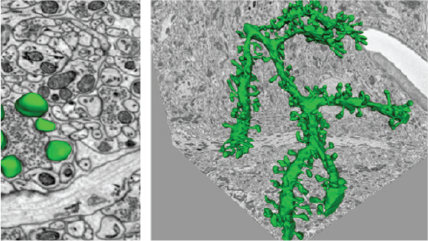
3View®システムにより生成されたマウス小脳の500枚の連続画像を含む、15,625 μm³ (25 x 25 x 25 μm)の三次元データセットを使用した樹状突起の三次元再構築像。樹状突起構造(緑色)、ボタン(黄色)、および小胞(赤色)。挿入画像は左上から時計回りに小脳の共焦点像、三次元モデルにレンダリングしたワイヤフレームトレース、超解像度の樹状突起棘モデルとシナプス、ワイヤフレームトレースを示す画像。
細胞生物学:共焦点の補完
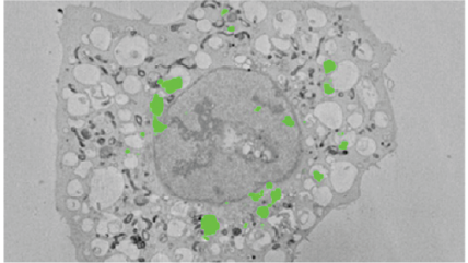
グリッド入りガラスボトムカバーガラスシャーレ上で培養されたLC-GFPを安定的に示しているヒーラ細胞の画像。無血清培地で2時間、飢餓状態にし、対象の細胞を共焦点顕微鏡法で識別したもの。細胞は電子顕微鏡用にin situで処理し、カバーガラスはフッ化水素酸を使用してエポキシ樹脂から溶解しています。細胞は樹脂ブロック内と、3Viewシステムで作成された連続画像内に観察されています。
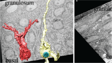
細胞生物学:発生
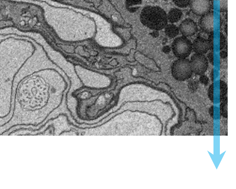
左上:高解像度、マウスの腎臓、1.5 nmピクセルで取得した8192 x 8192ピクセル画像。中央上:広視野、マウスの腎臓、80 nmピクセルで取得した8192 x 8192ピクセル画像。右:高圧凍結法で作製したC.エレガンス、25 nmピクセルで取得した4096 x 4096ピクセル画像。試料はカリフォルニア大学(バークレー)のKent McDonald様のご厚意によるものです。左下:マウスの坐骨神経、5 nmピクセルで取得した2048 x 2048ピクセル画像。中央下:マウスの坐骨神経軸索の三次元視覚化画像。
材料科学
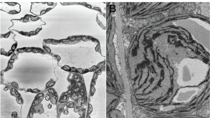
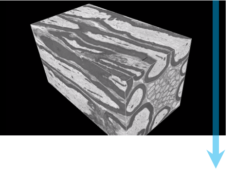
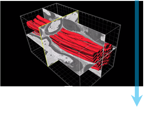
左上および左下:3Viewシステムで作成したアルミニウム表面の陽極酸化被覆の画像。中央:3Viewシステムで作成したアルミニウム合金とマンガン粒子の三次元可視化画像。3Dデータセットには、ピクセルサイズ15 nm、切片厚さ15 nmの1024 x 1024連続画像1,000枚が含まれています。3Dモデルは、3D可視化プラグインを使用してDigitalMicrograph®で作成されたものです。
シリアルブロックフェイスイメージングのワークフロー
|
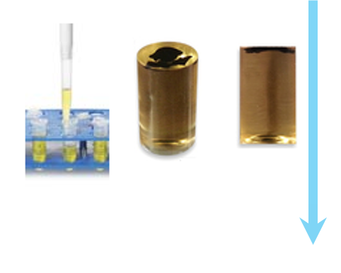
ステップ1:試料作製 通常、生物試料は固定し、染色液で染色し、安定に保持するため樹脂に埋め込みます。 |
![]() |
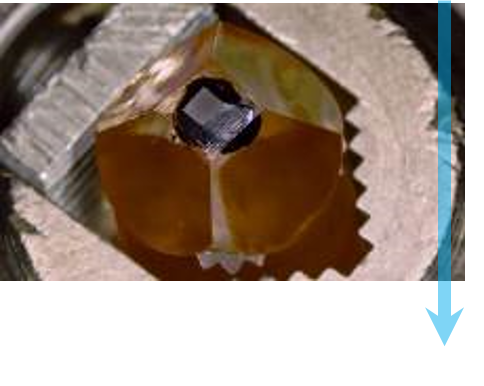
ステップ2:試料マウントとSEMへの移送 試料は必要な寸法に合わせてトリミングし、アルミニウムピンに載せて固定し、任意で金薄スパッタコーティングを行います。試料ピンを3Viewシステムに固定し、ダイヤモンドナイフを接触させ、ドアを閉めて排気します。 |
![]() |
ステップ3:イメージングの最適化 標準的なSEM条件に従い、ただし、Z方向の影響を考慮して、適切なビーム条件を選択します。画像の解像度、視野、取得時間が目的の条件になるように、倍率、ピクセル数、およびドウェルタイムを選択します。 |
![]() |
ステップ4:自動像観察 選択したビーム条件で連続画像が取得されます。各画像を取得する前に、ナイフで試料の表面層が切り取られます。 |
![]() |
ステップ5:解析 画像は三次元データセットを形成するためにスタックされ、このデータセットをDigitalMicrograph®またはサードパーティ製のソフトウェアで処理して表示できます。この時点で、セグメンテーションと定量化が可能になります。 |
Gatan Microscopy Suiteとしても知られるDigitalMicrographソフトウェア。デジタルカメラの制御と共に、電子線トモグラフィ、その場観察、スペクトラムイメージング、ディフラクションイメージング、その他多くのアプリケーションをサポート。
3D-CLEM reveals that a major portion of mitotic chromosomes is not chromatin
Booth, D. G.; Beckett, A. J.; Molina, O.; Samejima, I.; Masumoto, H.; Kouprina, N.; Larionov, V.; Prior, I. A.; Earnshaw, W. C.
Holcomb, P. S.; Hoffpauir, B. K.; Hoyson, M. C.; Jackson, D. R.; Deerinck, T. J.; Marrs, G. S.; Dehoff, M.; Wu, J.; Ellisman, M. H.; Spirou, G. A.
High-throughput 3D visualization of large volumes at high resolution by 3View
Dohnalkova, A.; Kennedy, D.; Mancuso, J.; Marshall, M.; Mainwaring, P.; Fredrickson, J.
Lead asparate, an en bloc contrast stain particularly useful for ultrastructural enzymology
Walton, J.
Morphology of phagophore precursors by correlative light-electron microscopy
Gudmundsson, S. R.; Kallio, K. A.; Vihinen, H.; Jokitalo, E.; Ktistakis, N.; Eskelinen, E. L.
Tanaka, T.; Ohno, N.; Osanai, Y.; Saitoh, S.; Thai, T. Q.; Nishimura, K.; Shinjo, T.; Takemura, S.; Tatsumi, K.; Wanaka, A.
Mitochondrial morphology and function: two for the price of one!
Faitg, J.; Davey, T.; Turnbull, D. M.; White, K.; Vincent, A. E.
Ultrastructural variability of the juxtacanalicular tissue along the inner wall of Schlemm’s canal
Koudouna, E.; Young, R. D.; Overby, D. R.; Ueno, M.; Kinoshita, S.; Knupp, C.; Quantock, A. J.
Hashimoto, T.; Thompson, G. E.; Zhou, X.; Withers, P. J.
The biosynthesis of infrared-emitting quantum dots in allium fistulosum
Green, M.; Haigh, S. J.; Lewis, E. A.; Sandiford, L.; Burkitt-Gray, M.; Fleck, R.; Vizcay-Barrena, G.; Jensen, L.; Mirzai, H.; Curry, R. J.; Dailey, L. -A.
Borrett, S.; Hughes, L.
Kreshuk, A.; Walecki, R.; Koethe, U.; Gierthmuehlen, M.; Plachta, D.; Genoud, C.; Haastert-Talini, K.; Hamprecht, F. A.
Froud, K. E.; Wong, A. C. Y.; Cederholm, J. M. E.; Klugmann, M.; Sandow, S. L.; Julien, J. -P.; Ryan, A. F.; Housley, G. D.
Challenges of microtome-based serial block-face scanning electron microscopy in neuroscience
Wanner, A. A.; Kirschmann, M. A.; Genoud, C.
High-resolution whole-brain staining for electron microscopic circuit reconstruction
Mikula, S.; Denk, W.
Comparison and combination of imaging techniques for three dimensional analysis of electrical trees
Schurch, R.; Rowland, S.M.; Bradley, R.S.; Withers, P.J.
The Lowe syndrome protein OCRL1 is required for endocytosis in the zebrafish pronephric tubule
Oltrabella, F.; Pietka, G.; Ramirez, I. B.-R.; Mironov, A.; Starborg, T.; Drummond, I. A.; Hinchliffe, K. A.; Lowe, M.
Starborg, T.; Kadler, K. E.
Hondow, N.; Brown, M. R.; Starborg, T.; Monteith, A. G.; Brydson, R.; Summers, H. D.; Rees, P.; Brown, A.
Three-dimensional architecture of podocytes revealed by block-face scanning electron microscopy
Ichimura, K.; Miyazaki, N.; Sadayama, S.; Murata, K.; Koike, M.; Nakamura, K. -I.; Ohta, K.; Sakai, T.
Analysis of primary cilia in the developing mouse brain
Paridaen, J. T. M. L.; Huttner, W. B.; Wilsch-Bräuninger, M.
How to bury the dead: elimination of apoptotic hair cells from the hearing organ of the mouse
Anttonen, T.; Belevich, I.; Kirjavainen, A.; Laos, M.; Brakebusch, C.; Jokitalo, E.; Pirvola, U.
Developing 3D SEM in a broad biological context
Kremer, A.; Lippens, S.; Bartunkova, S.; Asselbergh, B.; Blanpain, C.; Fendrych, M.; Goossens, A.; Holt, M.; Janessens, S.; Krols, M.; Larsimont, J. -C.; Mc Guire, C.; Nowack, M. K.; Saelens, X.; Schertel, A.; Schepens, B.; Slezak, M.; Timmerman, V.; Theunis, C.; Van Brempt, R.; Visser, Y.; Guerin, C. J.
Application of FEA to image-based models of electrical trees with uniform conductivity
Rowland, S. M.; Schurch, R.; Pattouras, M.; Li, Q.
RBM3 mediates structural plasticity and protective effects of cooling in neurodegeneration
Peretti, D.; Bastide, A.; Radford, H.; Verity, N.; Molloy, C.; Martin, M. G.; Moreno, J. A.; Steinert, J. R.; Smith, T.; Dinsdale, D.; Willis, A. E.; Mallucci, G. R.
Quantitative analysis of mouse pancreatic islet architecture by serial block-face SEM
Pfeifer, C. R.; Shomorony, A.; Aronova, M. A.; Zhang, G.; Cai, T.; Xu, H.; Notkins, A. L.; Leapman, R. D.
Advanced imaging and tissue engineering of the human limbal epithelial stem cell niche
Massie, I.; Dziasko, M.; Kureshi, A.; Levis, H. J.; Morgan, L.; Neale, M.; Sheth, R.; Tovell, V. E.; Vernon, A. J.; Funderburgh, J. L.; Daniels, J. T.
Kempen, P. J.; Kircher, M. F.; de la Zerda, A.; Zavaleta, C. L.; Jokerst, J. V.; Mellinghoff, I. K.; Gambhir, S. S.; Sinclair, R.
Pinali, C.; Holt, C. M.; Bennett, H. J.; Davenport, J. B.; Walker, R.; Murfitt, L.; Allan, L. J.; Malik, N.; Kitmitto, A.
Automated analysis of spine dynamics on live CA1 pyramidal cells
Blumer, C.; Vivien, C.; Genoud, C.; Perez-Alvarez, A.; Wiegert, J. S.; Vetter, T.; Oertner, T. G.
Automated analysis of spine dynamics on live CA1 pyramidal cells
Blumer, C.; Vivien, C.; Genoud, G.; Perez-Alvarez, A.; Wiegert, J. S.; Vetter, T.; Oertner, T. G.
Ju, W. K.; Kim, K. Y.; Noh, Y. H.; Hoshijima, M.; Lukas, T. J.; Ellisman, M. H.; Weinreb, R. N.; Perkins, G. A.
Finite element analysis of the pressure-induced deformation of Schlemm’s canal endothelial cells
Vargas-Pinto, R.; Lai, J.; Gong, H.; Ethier, C. R.; Johnson, M.
Serial block face-scanning electron microscopy: a method to study retinal degenerative phenotypes
Mustafi, D.; Kikano, S.; Palczewski, K.
Serial block face-scanning electron microscopy: A method to study retinal degenerative phenotypes
Mustafi, D.; Kikano, S.; Palczewski, K.
Staining and embedding of human chromosomes for 3-d serial block-face scanning electron microscopy
Yusuf, M.; Chen, B.; Hashimoto, T.; Estandarte, A. K.; Thompson, G.; Robinson, I.
Three-dimensional reconstruction of skeletal muscle extracellular matrix ultrastructure
Gillies, A. R.; Bushong, E. A.; Deerinck, T. J.; Ellisman, M. H.; Lieber, R. L.
The versatile electron microscope: An ultrastructural overview of autophagy
Biazik, J.; Vihinen, H.; Anwar, T.; Jokitalo, E.; Eskelinen, E.
Mission (im)possible – mapping the brain becomes a reality
Eberle, A. L.; Selchow, O.; Thaler, M.; Zeidler, D.; Kirmse, R.
Dynein light intermediate chains maintain spindle bipolarity by functioning in centriole cohesion
Jones, L. A.; Villemant, C.; Starborg, T.; Salter, A.; Goddard, G.; Ruane, P.; Woodman, P. G.; Papalopulu, N.; Woolner, S.; Allan, V. J.
Urwyler, O.; Izadifar, A.; Dascenco, D.; Petrovic, M.; He, H.; Ayaz, D.; Kremer, A.; Lippens, S.; Baatsen, P.; Guérin, C. J.; Schmucker, D.
Pinali, C.; Kitmitto, A.
En bloc staining with hydroquinone treatment for block face imaging
Togo, A.; Ohta, K.; Higashi, R.; Nakamura, K.
Three dimensional imaging of electrical trees in micro and nano-filled epoxy resin
Schurch, R.; Rowland, S. M.; Bradley, R. S.; Hashimoto, T.; Thompson, G. E.; Withers, P. J.
Three dimensional imaging of electrical trees in micro and nano-filled epoxy resin
Schurch, R.; Rowland, S.M.; Bradley, R.S.; Hashimoto, T.; Thompson, G.E.; Withers, P.J.
Finite-size effects in the 3D reconstruction and morphological analysis of porous polymers
Müllner, T.; Zankel, A.; Svec, F.; Tallarek, U.
Salloum, R. H.; Chen, G.; Velet, L.; Manzoor, N. F.; Elkin, R.; Kidd, GJ.; Coughlin, J.; Yurosko, C.; Bou-Anak, S.; Azadi, S.; Gohlsch, S.; Schneider, H.; Kaltenbach, J.A.
Salloum, R. H.; Chen, G.; Velet, L.; Manzoor, N. F.; Elkin, R.; Kidd, G. J.; Coughlin, J.; Yurosko, C.; Bou-Anak, S.; Azadi, S.; Gohlsch, S.; Schneider, H.; Kaltenbach, J.A.
A quantitative three-dimensional in situ study of a short fatigue crack in a magnesium alloy
Marrow, T. J.; Mostafavi, M.; Hashimoto, T.; Thompson, G. E.
A quantitative three-dimensional in situ study of a short fatigue crack in a magnesium alloy
Marrow, T. J.; Mostafavi, M.; Hashimoto, T.; Thompson, G. E.
Rhinomonas nottbecki n. sp. (cryptomonadales) and molecular phylogeny of the family pyrenomonadaceae
Majaneva, M.; Remonen, I.; Rintala, J. M.; Belevich, I.; Kremp, A.; Setälä, O.; Jokitalo, E.; Blomster, J.
Arabidopsis NAC45/86 direct sieve element morphogenesis culminating in enucleation
Furuta, K. M.; Yadav, S. R.; Lehesranta, S.; Belevich, I.; Miyashima, S.; Heo, J.; Vatén, A.; Lindgren, O.; De Rybel, B.; Van Isterdael, G.; Somervuo, P.; Lichtenberger, R.; Rocha, R.; Thitamadee, S.; Tähtiharju, S.; Auvinen, P.; Beeckman, T.; Jokitalo, E.; Helariutta, Y.
Tsai, W. T.; Hassan, A.; Sarkar, P.; Correa, J.; Metlagel, Z.; Jorgens, D. M.; Auer, M.
Busskamp, V.; Krol, J.; Nelidova, D.; Daum, J.; Szikra, T.; Tsuda, B.; Jüttner, J.; Farrow, K.; Scherf, B. G.; Alvarez, C. P.; Genoud, C.; Sothilingam, V.; Tanimoto, N.; Stadler, M.; Seeliger, M.; Stoffel, M.; Filipowicz, W.; Roska, B.
Miyazaki, N.; Esaki, M.; Ogura, T.; Murata, K.
Heras-Bautista, C. O.; Katsen-Globa, A.; Schloerer, N. E.; Dieluweit, S.; El Aziz OM, A.; Peinkofer, G.; Attia, W. A.; Khalil, M.; Brockmeier, K.; Hescheler, J.; Pfannkuche, K.
Miyazaki, N.; Esaki, M.; Ogura, T.; Murata, K.
Differential roles of microglia and monocytes in the inflamed central nervous system
Yamasaki, R.; Lu, H.; Butovsky, O.; Ohno, N.; Rietsch, A.M.; Cialic, R.; Wu, P. M.; Doykan, C. E.; Lin, J.; Cotleur, A. C.; Kidd, G.; Zorlu, M. M.; Sun, N.; Hu, W.; Liu, L.; Lee, J. C.; Taylor, S. E.; Uehlein, L.; Dixon, D.; Gu, J.; Floruta, C. M.; Zhu, M.; Charo, I. F.; Weiner, H. L.; Ransohoff, R. M.
Mitochondrial immobilization mediated by syntaphilin facilitates survival of demyelinated axons
Ohno, N.; Chiang, H.; Mahad, D. J.; Kidd, G. J.; Liu, L.; Ransohoff, R. M.; Sheng, Z. H.; Komuro, H. Trapp, B. D.
Cooperrider, J.; Furmaga, H.; Plow, E.; Park, H. J.; Chen, Z.; Kidd, G.; Baker, K. B.; Gale, J. T.; Machado, A. G.
Hughes, A. E.; Trinchi, A.; Chen, F. F.; Yang, Y. S.; Cole, I. S.; Sellaiyan, S.; Carr, J.; Lee, P. D.; Thompson, G. E.; Xiao, T. Q.
Serial sectioning methods for 3D investigations in materials science
Zankel, A.; Wagner, J.; Poelt, P.
Lipke, E.; Hörnschemeyer, T.; Pakzad, A.; Booth, C. R.; Michalik, P.
Chen, B.; Hashimoto, T.; Vergeer, F.; Burgess, A.; Thompson, G.; Robinson, I.
Pinali, C.; Bennett, H. J.;Davenport, J. B.; Caldwell, J. L.; Trafford, A. W.; Kitmitto, A.
Lipke, E.; Hörnschemeyer, T.; Pakzad, A.; Booth, C. R.; Michalik, P.
Pinali, C.; Bennett, H. J.; Davenport, J. B.; Caldwell, J. L.; Trafford, A. W.; Kitmitto, A.
Transcellular degradation of axonal mitochondria
Davis, C. O.; Kim, K.; Bushong, E. A.; Mills, E. A.; Boassa, D.; Shih, T.; Kinebuchi, M.; Phan, S.; Zhou, Y.; Bihlmeyer, N. A.; Nguyen, J. V.; Jin, Y.; Ellisman, M. H.; Marsh-Armstrong, N.
Revelation of intertwining organic and inorganic fractal structures in polymer coatings
Hughes, A. E.; Trinchi, A.; Chen, F. F.; Yang, Y. S.; Cole, I. S.; Sellaiyan, S.; Carr, J.; Lee, P. D.; Thompson, G. E.; Xiao, T. Q.
Dziasko, M. A.; Armer, H. E.; Levis, H. J.; Shortt, A. J.; Tuft, S.; Daniels, J. T.
Wu, J.; Bakerink, K. J.; Evangelista, M. E.; Thomas, G. H.
ER sheet persistence is coupled to myosin 1c–regulated dynamic actin filament arrays
Joensuu, M.; Belevich, I.; Rämö, O.; Nevzorov, I.; Vihinen, H.; Puhka, M.; Witkos, T. M.; Lowe, M.; Vartiainen, M. K.; Jokitalo, E.
Gillies, A.; Lieber, R.
Serial block face scanning electron microscopy-the future of cell ultrastructure imaging
Hughes, L.; Hawes, C.; Monteith, S.; Vaughan, S.
An enteroendocrine cell-enteric glia connection revealed by 3D electron microscopy
Bohórquez, D. V.; Samsa, L. A.; Roholt, A.; Medicetty, S.; Chandra, R.;Liddle, R. A.
Pingel, J.; Lu, Y.; Starborg, T.; Fredberg, U.; Langberg, H.; Nedergaard, A.; Weis, M.; Eyre, D.; Kjaer, M.; Kadler, K. E.
Resolution of the three dimensional structure of components of the glomerular filtration barrier
Arkill, K. P; Qvortrup, K.; Starborg, T.; Mantell, J. M.; Knupp, C.; Michel, C. C.; Harper, S. J.; Salmon, A. H. J.; Squire, J. M.; Bates, D. O.; Neal, C. R.
Three-dimensional aspects of matrix assembly by cells in the developing cornea
Young, R. D.; Knupp, C.; Pinali, C.; Png, K. M. Y.; Ralphs, J. R.; Bushby, A. J.; Starborg,T.; Kadler, K. E.; Quantock, A. J.
Wilke, S. A.; Raam, T.; Antonios, J. K.; Bushong, E. A.; Koo, E. H.; Ellisman, M. H.; Ghosh, A.
Resolution of three dimensional structure of components of the glomerular filtration barrier
Arkill, K. P.; Qvortrup, K.; Starborg, T.; Mantell, J. M.; Knupp, C.; Michel, C. C.; Harper, S. J.; Salmon, A. H. J.; Squire, J. M.; Bates, D. O.; Neal, C. R.
Exploring the third dimension: Volume electron microscopy comes of age
Peddie, C. J.; Collinson, L. M.
Hughes, L.; Towers, K.; Starborg, T.; Gull, K.; Vaughan, S.
The protein quality control system manages plant defence compound synthesis
Pollier, J.; Moses, T.; González-Guzmán, M.; De Geyter, N.; Lippens, S.; Bossche, R. V.; Marhavý, P.; Kremer, A.; Morreel, K.; Guérin, C. J.; Tava, A.; Oleszek, W.; Thevelein, J. M.; Campos, N.; Goormachtig, S.; Goossens, A.
Nonmuscle myosin II powered transport of newly formed collagen fibrils at the plasma membrane
Kalson, N. S.; Starborg, T.; Lu, Y.; Mironov, A.; Humphries, S. M.; Holmes, D. F.; Kadler, K. E.
Widespread mitochondrial depletion via mitiphagy does not compromise necroptosis
Tait, S. W.; Oberst, A.; Quarato, G.; Milasta, S.; Haller, M.; Wang, R.; Karvela, M.; Ichim, G.; Yatim, N.; Albert, M. L.; Kidd, G.; Wakefield, R.; Frase, S.; Krautwald, S.; Linkermann, A.; Green, D. R.
Pinali, C.; Bennett, H.; Davenport, J. B.; Trafford, A. W.; Kitmitto, A.
Kuwajima, M.; Spacek, J.; Harris, K. M.
Trueman, A.; Knight, S.; Colwell, J.; Hashimoto, T.; Carr, J.; Skeldon, P.; Thompson, G.
Connectomic reconstruction of the inner plexiform layer in the mouse retina
Helmstaedter, M.; Briggman, K. L.; Turaga, S. C.; Jain, V.; Seung, H. S.; Denk, W.
Dwyer, L.; Robson, J.; da Fonseca, J. Q.; Kamp, N.; Hashimoto, T.; Thompson, G. E.
Construction of a polarized neuron
Holcomb, P. S.; Deerinck, T. J.; Ellisman, M. H.; Spirou, G. A.
Hashimoto, T.; Thompson, G. E.; Curioni, M.; Zhou, X. R.; Skeldon, P.
Three dimensional electron microscopy of cellular organelles by serial block face SEM and ET
Vihinen, H.; Belevich, I.; Jokitalo, E.
Wong, J.; Baddeley, D.; Bushong, E. A.; Yu, Z.; Ellisman, M. H.; Hoshijima, M.; Soeller, C.
Functional development of the olfactory system in zebrafish
Miyasaka, N.; Wanner, A. A.; Li, J.; Mack-Bucher, J.; Genoud, C.; Yoshihara, Y.; Friedrich, R. W.
BMP signaling specifies the development of a large and fast CNS synapse
Xiao, L.; Michalski, N.; Kronander, E.; Gjoni, E.; Genoud, C.; Knott, G.; Schneggenburger, R.
DP2: Distributed 3D image segmentation using micro-labor workforce
Giuly, R. J.; Kim, K. Y.; Ellisman, M. H.
Shigetomi, E.; Bushong, E. A.; Haustein, M. D.; Tong, X.; Jackson-Weaver, O.; Kracun, S.; Xu, J.; Sofroniew, M. V.; Ellisman, M. H.; Khakh, B. S.
Analyzing the structure and function of neuronal circuits in zebrafish
Friedrich, R. W.; Genoud, C.; Wanner, A. A.
WDR81 is necessary for purkinje and photoreceptor cell survival
Traka, M.; Millen, K. J.; Collins, D.; Elbaz, B.; Kidd, G. J.; Gomez, C. M.; Popko, B.
Large-scale automatic reconstruction of neuronal processes from electron microscopy images
Kaynig, V.; Vazquez-Reina, A.; Knowles-Barley, S.; Roberts, M.; Jones, T. R.; Kasthuri, N.; Miller, E.; Lichtman, J.; Pfister, H.
Boassa, D.; Berlanga, M. L.; Yang, M. A.; Terada, M.; Hu, J.; Bushong, E. A.; Hwang, M.; Masliah, E.; George, J. M.; Ellisman, M. H.
Revealing the three dimensional internal structure of aluminium alloys
Thompson, G. E.; Hashimoto, T.; Zhong, X. L.; Curioni, M.; Zhou, X.; Skeldon, P.; Withers, P. J.; Carr, J. A.; Monteith, A. G.
Jurrus, E.; Watanabe, S.; Giuly, R. J.; Paiva, A. R.; Ellisman, M. H.; Jorgensen, E. M.; Tasdizen, T.
Wilke, S. A.; Antonios, J. K.; Bushong, E. A.; Badkoobehi, A.; Malek, E.; Hwang, M.; Terada, M.; Ellisman, M. H.; Ghosh, A.
Autosomal recessive retinitis pigmentosa E150K opsin mice exhibit photoreceptor disorganization
Zhang, N.; Kolesnikov, A. V.; Jastrzebska, B.; Mustafi, D.; Sawada, O.; Maeda, T.; Genoud, C.; Engel, A.; Kefalov, V. J.; Palczewski, K.
Starborg, T.; Kalson, N. S.; Lu, Y.; Mironov, A.; Cootes, T. F.; Holmes, D. F.; Kadler, K. E.
Paridaen, J. T. M. L.; Wilsch-Bräuninger, M.; Huttneremail, W. B.
Three-dimensional structure analysis and percolation properties of a barrier marine coating
Chen, B.; Guizar-Sicairos,M.; Xiong,G.; Shemilt, L.; Diaz, A.; Nutter, J.; Burdet,N.; Huo,S.; Mancuso, J.; Monteith,A.; Vergeer,F.; Burgess,A.; Robinson, I.
Anttonen, T.; Kirjavainen, A.; Belevich, I.; Laos, M.; Richardson, W. D.; Jokitalo, E.; Brakebusch, C.; Pirvola, U.
Müllner, T.; Zankel, A.; Mayrhofer, C.; Reingruber, H.; Höltzel, A.; Lv, Y.; Svec, F.; Tallarek, U.
Investigation of dealloying by ultra-high-resolution nanotomography
Hashimoto, T.; Curioni, M.; Zhou, X.; Mancuso, J.; Skeldon, P.; Thompson, G. E.
Schwartz, C. L.; Heumann, J. M.; Dawson, S. C.; Hoenger,A.
Prdm3 and Prdm16 are H3K9me1 methyltransferases required for mammalian heterochromatin integrity
Pinheiro, I.; Margueron, R.; Shukeir, N.; Eisold, M.; Fritzsch, C.; Richter, F. M.; Mittler, G.; Genoud, C.; Goyama, S.; Kurokawa, M.; Son, J.; Reinberg, D.; Lachner, M.; Jenuwein, T.
Puhka, M.; Joensuu, M.; Vihinen, H.; Belevich, I.; Jokitalo, E.
Zhuravleva, E.; Gut, H.; Hynx, D.; Marcellin, D.; Bleck, C. K.; Genoud, C.; Cron, P.; Keusch, J. J.; Dummler, B.; Esposti, M. D.; Hemmings, B. A.
Azeloglu, E.; Stothers, M.; Deerinck, T. J.; Falkenberg, C.; Chen, Y.; He, J. C.; Hone, J. C.; Leslie, L. M.; Ellisman, M. H.; Iyengar, R.
Extrusion of misfolded and aggregated proteins--a protective strategy of aging neurons?
Doehner, J.; Genoud, C.; Imhof, C.; Krstic, D.; Knuesel, I.
3D segmentation of SBFSEM images of neuropil by a graphic model over supervoxel boundaries
Andres, B.; Koethe, U.; Kroeger, T.; Helmstaedter, M.; Briggman, K. L.; Denk, W.; Hamprecht, F. A.
Differential modulation of retinal degeneration by Ccl2 and Cx3cr1 chemokine signalling
Luhmann, U. F.; Lange, C. A.; Robbie, S.; Munro, P. M.; Cowing, J. A.; Armer, H. E.; Luong, V.; Carvalho, L. S.; MacLaren, R. E.; Fitzke, F. W.; Bainbridge, J.W.; Ali, R. R.
Beam deceleration for block-face scanning electron microscopy of embedded biological tissue
Ohta, K.; Sadayama, S.; Togo, A.; Higashi, R.; Tanoue, R.; Nakamura, K.
Multivesicular exocytosis in rat pancreatic beta cells
Hoppa, M. B.; Jones, E.; Karanauskaite, J.; Ramracheya, R.; Braun, M.; Collins, S. C.; Zhang, Q.; Clark, A.; Eliasson, L.; Genoud, C.; Macdonald, P. E.; Monteith, A. G.; Barg, S.; Galvanovskis, J.; Rorsman, P.
Structural neurobiology: Missing link to a mechanistic understanding of neural computation
Denk, W.; Briggman, K. L.; Helmstaedter, M.
Giuly, R. J.; Martone, M. E.; Ellisman, M. H.
Volume electron microscopy for neuronal circuit reconstruction
Briggman, K. L.; Bock, D. D.
Yan, J.; Walker, C. G.; O'sullivan, M. J.; Bushong, E. A.; Ellisman, M. H.; Hoshijima, M.; Rajagopal, V.
High contrast en bloc staining of neuronal tissue for field emission scanning electron microscopy
Tapia, J. C.; Kasthuri, N.; Hayworth, K. J.; Schalek, R.; Lichtman, J. W.; Smith, S. J.; Buchanan, J.
Modern electron microscopy methods for C. elegans
Hall, D. H.; Hartwieg, E.; Nguyen, K. C.
Ou, H. D.; Kwiatkowski, W.; Deerinck, T. J.; Noske, A.; Blain, K. Y.; Land, H. S.; Soria, C.; Powers, C. J.; May, A. P.; Shu, X.; Tsien, R. Y.; Fitzpatrick, J. A. J.; Long, J. A.; Ellisman, M. H.; Choe, S.; O'Sheaemail, C. C.
Mustafi, D.; Kevany, B. M.; Genoud, C.; Okano, K.; Cideciyan, A. V.; Sumaroka, A.; Roman, A. J.; Jacobson, S. G.; Engel, A.; Adams, M. D.; Palczewski, K.
Motskin, M.; Müller, K. H.; Genoud, C.; Monteith, A. G.; Skepper, J. N.
Kalson, N. S.; Holmes, D. F.; Herchenhan, A.; Lu, Y.; Starborg, T.; Kadler, K. E.
Wong, J. I. S.; Baddeley, D.; Bushong, E. A.; Ellisman, M.; Hoshijima, M.; Soeller, C.
Fast extraction of neuron morphologies from large-scale SBFSEM image stacks
Lang, S.; Drouvelis, P.; Tafaj, E.; Bastian, P.; Sakmann, B.
Mun, J. Y.; Jeong, S. Y.; Kim, J. H.; Han, S. S.; Kim, I. H.
Basement membrane changes in capillaries of the ageing human retina
Powner, M. B.; Scott, A.; Zhu, M.; Munro, P. M.; Foss, A. J.; Hageman, G. S.; Gillies, M. C.; Fruttiger, M.
Cadherin-9 regulates synapse-specific differentiation in the developing hippocampus
Williams, M. E.; Wilke, S. A.; Daggett, A.; Davis, E.; Otto, S.; Ravi, D.; Ripley, B.; Bushong, E. A.; Ellisman, M. H.; Klein, G.; Ghosh, A.
High-accuracy neurite reconstruction for high-throughput neuroanatomy
Helmstaedter, M.; Briggman, K. L.; Denk, W.
Role of elastin anisotropy in structural strain energy functions of arterial tissue
Rezakhaniha, R.; Fonck, E.; Genoud, C.; Stergiopulos, N.
Modeling of flow in a polymeric chromatographic monolith
Koku, H.; Maier, R. S.; Czymmek, K. J.; Schure, M. R.; Lenhoff, A. M.
Quantitative Characterisation of microfiltration membranes by 3D reconstruction
Reingruber, H.; Zankel, A.; Mayrhofer, C.; Poelt, P.
Shu, X.; Lev-Ram, V.; Deerinck, T. J.; Qi, T.; Ramko, E. B.; Davidson, M. W.; Jin, Y. Ellisman, M. H.; Tsien, R. Y.
Wiring specificity in the direction-selectivity circuit of the retina
Briggman, K. L.; Helmstaedter, M.; Denk, W.
Nguyen, J. V.; Soto, I.; Kim, K.; Bushong, E. A.; Oglesby, E.; Valiente-Soriano, F. J.; Yang, Z.; Davis, C. O.; Bedont, J. L.; Son, J. L.; Wei, J. O.; Buchman, V. L.; Zack, D. J.; Vidal-Sanz, M.; Ellisman, M. H.; Marsh-Armstrong, N.
Environmental scanning electron microscopy (ESEM)--a versatile tool in studying plants.
Stabentheiner, E.; Zankel A.; Pölt P.
Jurrus, E.; Paiva, A. R.; Watanabe, S.; Anderson, J. R.; Jones, B. W.; Whitaker, R. T.; Jorgensen, E. M.; Marc, R. E.; Tasdizen, T.
Reconstruction of a neuron from SBFSEM: tools, reliability, accuracy and efficiency
Drouvelis, P.; Kurz, T.; Bastian, P.; Sakmann, B.; Lang, S.
Deerinck, T. J.; Bushong, E. A.; Lev-Ram, V.; Shu, X.; Tsien, R. Y.; Ellisman, M. H.
Structure-function studies of blood and air capillaries in chicken lung using 3D electron microscopy
West, J. B.; Fu, Z.; Deerinck, T. J.; Mackey, M. R.; Obayashi, J. T.; Ellisman, M.H.
Imaging transient blood vessel fusion events in zebrafish by correlative volume electron microscopy
Armer, H. E. J.; Mariggi, G.; Png, K. M. Y.; Genoud, C.; Monteith, A. G.; Bushby, A. J.; Gerhardt, H.; Collinson, L. M.
Saetzler, K.; McCanny, P.; Rodriguez, E.P.; Horstmann, H.; Bruno, R.M.; Denk, W.
Axon tracking in serial block-face scanning electron microscopy
Jurrus, E.; Hardy, M.; Tasdizen, T.; Fletcher, P. T.; Koshevoy, P.; Chien, C. B.; Denk, W.; Whitaker, R.
Mishchenko, Y.
Three-dimensional reconstruction methods for Caenorhabditis elegans ultrastructure
Müller-Reichert, T.; Mancuso, J.; Lich, B.; McDonald, K.
Ultramicrotomy in the ESEM, a versatile method for materials and life sciences
Zankel, A.; Kraus, B.; Poelt, P.; Schaffer, M.; Ingolic, E.
Genoud, C.; Mancuso, J.; Monteith, S.; Kraus, B.
Contour-propagation algorithms for semi-automated reconstruction of neural processes
Macke, J. H.; Maack, N.; Gupta, R.; Denk, W.; Schölkopf, B.; Borst, A.
Segmentation of SBFSEM volume data of neural tissue by hierarchical classification
Andres, B.; Köthe, U.; Helmstaedter, M.; Denk, W.; Hamprecht, F. A.
Alpha-herpesvirus infection induces the formation of nuclear actin filaments
Feierbach, B.; Piccinotti, S.; Bisher, M.; Denk, W.; Enquist, L. W.
Serial block-face scanning electron microscopy to reconstruct three-dimensional tissue nanostructure
Denk, W.; Horstmann, H.