K3 IS Camera

The world’s first counting, high-speed, large-format camera for in-situ microscopy.

Advantages Research Spotlight Media Library Publications Resources Back to top
Advantages: 

K3® IS – The world’s only counting, high-speed, large-format camera for in-situ transmission electron microscopy (TEM). With an unprecedented temporal resolution, this true next-generation camera collects the ultimate in-situ data to extend the K3’s resolution revolution into material science.

Better

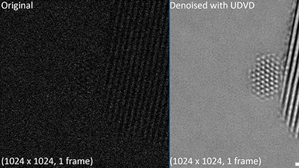
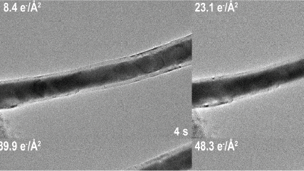
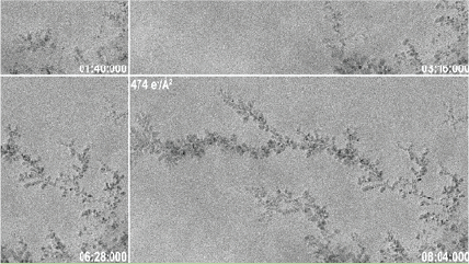
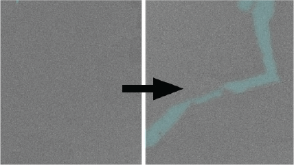
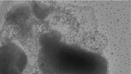
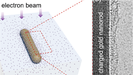
  • See your sample, not beam-induced artifacts
  • Capture the highest-quality, low-dose, in-situ video with the industry-leading DQE and sensitivity

Faster

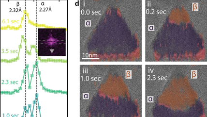
  • Count single electrons at unsurpassed speeds
    • 150 frames per second (fps) at full field of view
    • >3500 fps at 256 x 256 pixels
  • Shorten time to results with the market-leading Gatan Microscopy Suite® (GMS) in-situ processing utilities and free offline tools

Larger

  • Expand the field of view to 14- or 24-megapixels – Up to 1.65 times the size of the K2® IS camera
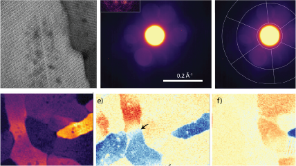
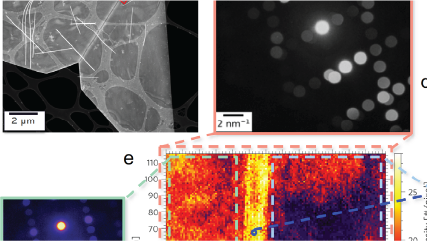
Research Spotlight

NUANCE

About the NUANCE Center

The Northwestern University's Atomic and Nanoscale Characterization Experimental Center  (...

The Center for Integrated Nanotechnologies

An Office of Science user facility jointly operated by Los Alamos and Sandia National Laboratories.

The Center for Integrated Nanotechnologies (CINT) offers...

Publications

2023

Kanomi, S.; Marubayashi, H.; Miyata, T.; Jinnai, H.

Microscopy and Microanalysis
2022

Pakzad, A.; dos Reis. R. D.

Resources:


Models 1026, 1027

Datasheet

K3 IS Camera

Applications

K3 IS camera for electron ptychography: Mapping oxygen in SrTiO3

Acquiring counted electron diffraction data without a beam stop with Gatan electron counting direct detectors

 In-situ lattice-resolution imaging of hydrogen absorption into nanoparticles

 In-situ observation of the annealing of Pt islands into branched Ru nanostructures to make single-atom catalysts

Imaging carbon nanoparticle agglomeration on MoS2 at a low dose rate

Imaging a lithium metal battery solid electrolyte interphase

Imaging of graphene at 200 kV using electron counting

Electric field mapping in 2D heterostructures using differential phase contrast

Magnetite nanoparticle orientation mapping from a 4D STEM dataset

Dynamic in-situ lithiation of NiS-filled carbon nanotubes

Electron counting 4D STEM studies of human tooth enamel

Grain boundary structure of two-dimensional tellurium revealed by 4D STEM

Observing beam-induced dendritic growth over two different timescales

Magnetite nanoparticle orientation mapping from a single low-dose transmission electron microscope image

Virtual (BF/DF) imaging reveals the position and concentration of precipitates in a Ni-W alloy

Electric field-induced structural dynamics in MoS2 observed using in-situ transmission electron microscopy

K3 IS: Low dose EM meets catalysis

Imaging discrete ions at a liquid-solid interface using low-dose cryo-electron microscopy (cryo-EM) and electron counting


Acknowledgment

Continuing our prosperous collaborations that built the K2, the K3 is the successful result of Peter Denes' team at Lawrence Berkeley National Laboratory and David Agard.

Back to top