The Center for Integrated Nanotechnologies
An Office of Science user facility jointly operated by Los Alamos and Sandia National Laboratories.
The Center for Integrated Nanotechnologies (CINT) offers world-leading scientific expertise and specialized capabilities to create, characterize, and integrate nanostructured materials at a range of length scales, from nano- to meso-scale. CINT helps scientists perform cutting-edge research in the areas of nanoscience and nanotechnology, providing FREE ACCESS for nonproprietary research. CINT facilities are open to the international research community, including government, academic, and industry researchers.
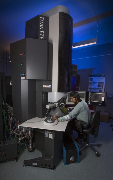
One of CINT’s core research areas is in-situ S/TEM experiments. Early capabilities focused on electrical and electrochemical property measurements of materials during high-resolution imaging. More recently, the staff has expanded the available techniques to include gaseous environments (ETEM), liquid-cell, mechanics, ion irradiation, laser ablation, cryogenic, heating, and air-free transfer. In-situ investigations have focused on coupled structure-property measurements on real materials, with the CINT user community expanding many of the in-situ method developments.
CINT Capability Highlights
Energy Storage: Solid-liquid interfacial reactions control the performance of many of today’s portable energy storage technologies. Understanding the mechanisms of ion transfer across these interfaces provides critical insight for engineering solutions to improve performance, such as artificial solid-electrolyte interphases. CINT can analyze these materials and systems using a variety of techniques.
-
Open-Cell In-Situ Electrochemistry: Individual nanowires or nanoparticles may be observed during reaction with charge-transfer ions (lithium, sodium, potassium, etc.)
-
Closed-Cell In-Situ Electrochemistry: CINT’s Electrochemical Discovery Platform is a custom-designed microelectromechanical system platform
-
Cryogenic FIB Lift-Out and Cryo-S/TEM: Intact investigations of electrode/electrolyte interfaces may be achieved by preparing an electrode-surface cross-section under inert transfer and cryogenic conditions in a focused ion beam (FIB)
-
Cryo-TEM: Investigations of electrolytes may be studied using plunge-freezing methods to rapidly cool a thin electrolyte layer, then image any structural features using TEM
-
Vacuum-Transfer S/TEM Holder: Allows for loading of a TEM grid in an Ar-filled glovebox for direct transfer into the S/TEM or ETEM
Soft Matter and Beam Sensitive Materials: CINT recently established a cryo-TEM suite dedicated to minimizing the electron dose for imaging materials and interfaces in their native hydrated (or solvated state). This provides an optimized characterization of sensitive materials across multiple length scales. Instruments included are a Talos dedicated cryo-TEM, a Scios 2 Dual-Beam SEM/FIB equipped with a Leica cryogenically-cooled stage, and a Vitrobot for reproducible and controlled vitrification of hydrated samples. Uniquely, CINT also provides Leica cryogenic-transfer equipment including a sample-prep station (VCM), high-vacuum transfer shuttle (VCT500), and a cryogenically-cooled sputter coater (ACE600). This robust and complete workflow allows for the preparation of frozen, electron-transparent lamellae that can be transferred to the TEM without ever leaving a cryogenic environment.
-
Talos L120C: Dedicated low dose, low kV TEM (user switchable from 20-120 kV) for imaging beam sensitive materials, and analyzing buried sample features and interfaces
-
Scios 2 Dual Beam Cryo-SEM/FIB: Contains a cryo-cooled SEM stage for surface analysis, analysis of buried interfaces, and serial sectioned 3D tomography of soft matter and nanomaterials in their native, hydrated state
-
Leica Transfer Equipment: Leica EM VCM enables users to easily put their cryogenic samples on holders under liquid nitrogen
Corrosion: Reactions at the surfaces of metals in contact with various solutions can cause massive structural deterioration, in the form of uniform, localized, and pitting corrosion. In-situ S/TEM has recently been proven as a method to investigate the initiation and propagation mechanisms of localized corrosion, with implications for progression mechanisms that cause pit formation.
-
S/TEM Experimental Methodology for Pre-, In-situ, and Post-Characterization: Using ACOM, energy-filtered TEM, EDS mapping, nanobeam diffraction, and high-resolution imaging, the metal sample may be mapped structurally and for the identification of compositional variations
-
Cryo-FIB/SEM/EDS/EBSD: Bulk corrosion samples with the corrosive medium intact can be studied using the Scios SEM with an attached Leica cryo-stage
Gas-solid Reactions: CINT offers a 300kV Titan G2 Environmental TEM which is monochromated, image-corrected, and equipped with a Gatan K2-IS camera. The gas cart provides high-level control over the inlet gas composition while minimizing contamination throughout the system. Many different studies may be conducted with an array of TEM holders. We also offer Protochips’ AXON software for minimizing image drift during in-situ reactions.
-
Catalysis: Gasses fed directly to the objective lens in the TEM include oxygen, hydrogen, nitrous oxide, methane, carbon monoxide, carbon dioxide, nitrogen, argon, and helium
-
Environmental Surface Reactions: Oxidation, reduction, annealing, and electrical break down mechanisms may be investigated
-
Hydrogenation: Coupled mechanical studies may be performed using a Hysitron PI-95 TEM holder
-
Vapor-Phase Corrosion: A new system has been installed to allow vapor introduction into the ETEM, including water for deliquescence corrosion studies
Publication Highlights
- Hattar, K.; Bufford, D. C.; Buller, D. L., Concurrent in situ ion irradiation transmission electron microscope. Nuclear Instruments and Methods in Physics Research Section B: Beam Interactions with Materials and Atoms 338 (2014): 56-65.
- Grosso, R.; Muccillo, E. N. S.; Muche, D. N. F.; Jawaharram, G. S.; Barr, C. M.; Monterrosa, A. M.; Castro, R. H. R.; Hattar, K.; Dillon, S. J., In situ transmission electron microscopy for ultrahigh temperature mechanical testing of ZrO2. Nano Letters (2020).
- Bufford, D. C., Stauffer, D.; Mook, W. M.; Syed Asif, S. A.; Boyce, B. L.; Hattar, K., High cycle fatigue in the transmission electron microscope. Nano Letters 16, no. 8 (2016): 4946-4953.
- Watt, J.; Huber, D. L.; Stewart, P. L., Soft matter and nanomaterials characterization by cryogenic transmission electron microscopy. MRS Bull. 2019, 44 (12), 942-948.
- Poerwoprajitno, A. R.; Gloag, L.; Benedetti, T. M.; Cheong, S.; Watt, J.; Huber, D. L.; Gooding, J. J.; Tilley, R. D., Formation of branched ruthenium nanoparticles for improved electrocatalysis of oxygen evolution reaction. Small 2019, 15 (17), e1804577.
- Watt, J.; Austin, M. J.; Simocko, C. K.; Pete, D. V.; Chavez, J.; Ammerman, L. M.; Huber, D. L., Formation of metal nanoparticles directly from bulk sources using ultrasound and application to e‐waste upcycling. Small 2018, 14 (17), 1703615.
- Hayden, S. C.; Chisholm, C.; Grudt, R. O.; Aguiar, J. A.; Mook, W. M.; Kotula, P. G.; Pilyugina, T. S.; Bufford, D.C.; Hattar, K.; Kucharski, T. J.; Taie, I. M.; Orstraat, M. L.; Jungjohann, K. L., Localized corrosion of low-carbon steel at the nanoscale, npj Materials Degradation 3(1), 1 (2019).
- Harrison, K.; Zavadil, K. R.; Hahn, N. T.; Meng, X.; Elam, J. W.; Leenheer, A.; Zhang, J. -G.; Jungjohann, K. L., Lithium self-discharge and its prevention: Direct visualization through in-situ electrochemical scanning transmission electron microscopy, ACS Nano 11, 11194 (2017).
- Xie, H.; Tan, X.; Luber, E. J.; Olsen, B.; Kalisvaart, W. P.; Jungjohann, K. L.; Mitlin, D.; Buriak, J. M., β-SnSb for Sodium ion battery anodes: Phase transformations responsible for enhanced cycling stability revealed by in-situ TEM. ACS Energy Letters 3(7), 1670 (2018).

