A Comprehensive In-Situ TEM Ecosystem from Gatan
Gatan’s continuously growing in-situ ecosystem is the most comprehensive, fully synchronized in-situ TEM ecosystem for multi-technique data collection, processing, and analysis. The latest Gatan TEM products and DigitalMicrograph make the entire in-situ experiment workflow easier, more powerful, and more flexible than ever before.
Au nanocrystal reorientation (video)
Video courtesy JEOL Japan and Gatan, Inc., US
This OneView camera video showcases step motion on the surface of a Au nanocrystal under the electron beam, however it is cropped to a smaller region of interest than the original image. During in-situ recordings, individual images at obtained at full resolution (4k x 4k) so you can create high resolution movies from the entire sample area or just a sub-region. Original Au nanocrystal reorientation image can be found in the OneView media library.
Sample: gold (Au) nanoparticles; beam energy: 300 kV; original image size: 4k x 4k; frame rate: 25 fps; binning x1
Rio camera for MicroED/3DED
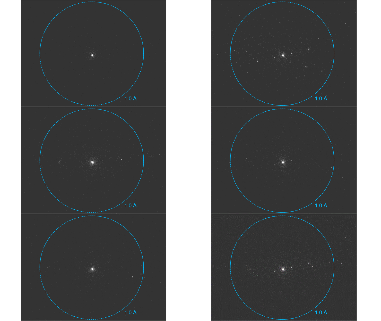
A three-dimensional electron diffraction dataset was collected from a biotin crystal with the Rio camera.
OneView camera + Latitude D for MicroED/3DED
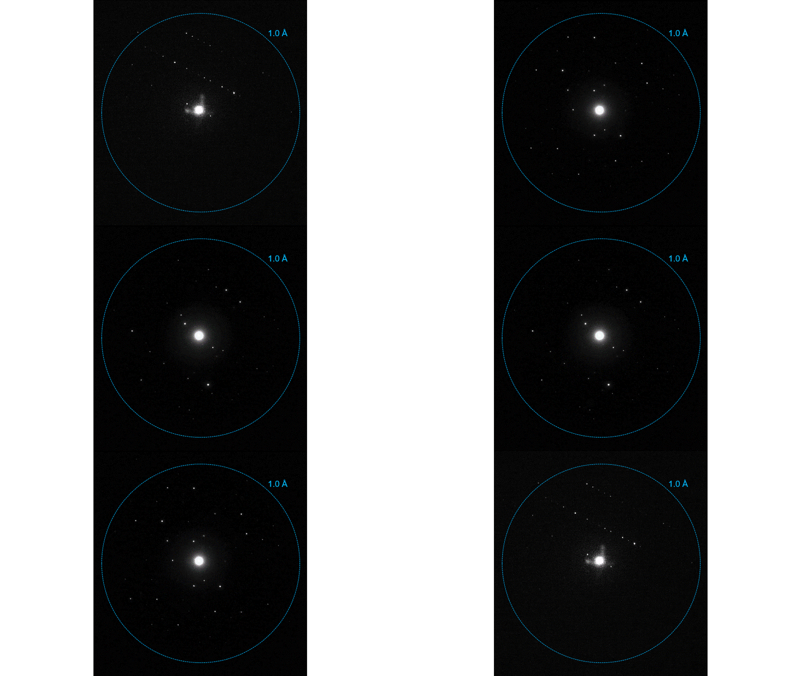
A three-dimensional electron diffraction dataset was collected from a ZSM-5 crystal collected with the OneView camera and Latitude D software. Diffraction patterns were recorded at 0.5° intervals collected over 100°. The cumulative dose was ∼2 e-/Å2.
Metro camera for MicroED/3DED
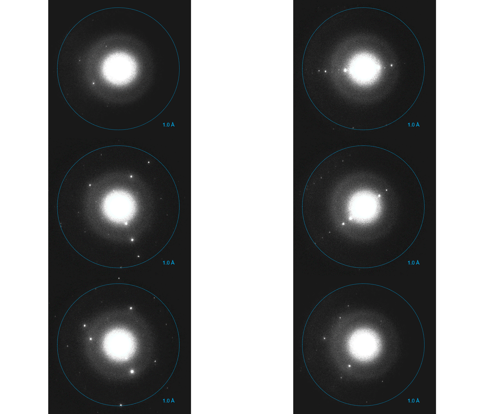
A three-dimensional electron diffraction dataset was collected with the Metro camera from a PPEA crystalline sample (courtesy of Dr. Tatiana Gorelik).
K3 camera for MicroED/3DED
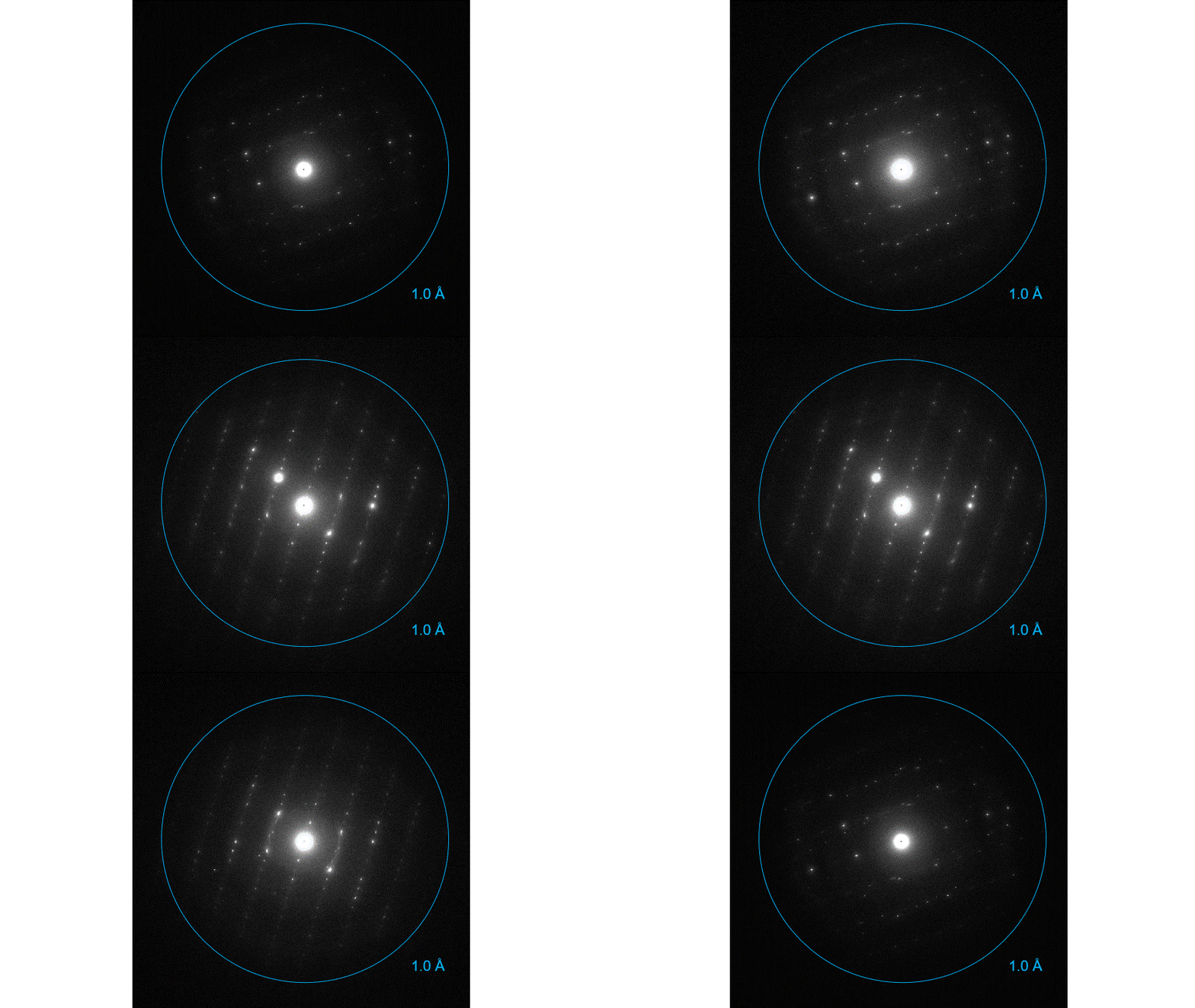
Counted three-dimensional electron diffraction dataset from a ZSM-5 crystal collected with the K3® camera. Diffraction patterns were recorded at 0.5° intervals collected over 10°. The cumulative dose was ∼0.5 e-/Å2
Stela camera for MicroED/3DED
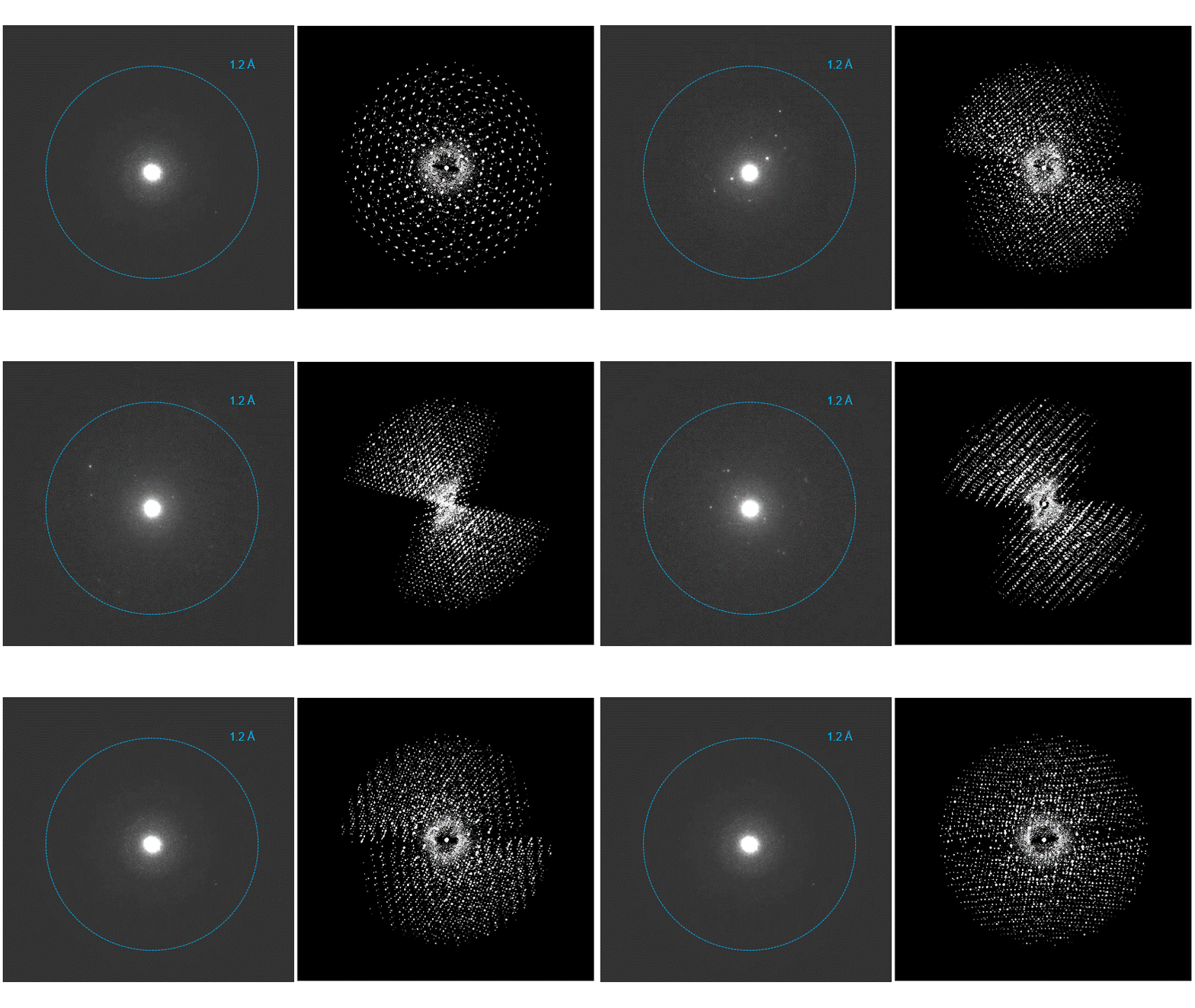
(left) A complete three-dimensional electron diffraction dataset from an acetaminophen microcrystal was acquired with a Stela® camera. (right) reciprocal space. Data was processed by Dr. Dos Reis at Northwestern University. Stela utilizes DECTRIS hybrid-pixel technology.
Latitude D workflow
Latitude D data collection workflow. (1) Start by specifying the Atlas size and click Capture to create an Atlas; (2) Choose a grid square; (3) Select one or multiple crystals; (4) Screen selected crystals in diffraction mode to check their quality; (5) Schedule MicroED/3DED data collection and walk away for the remainder of the acquisition.
Multimodal spectrum imaging at low kV with GIF Continuum K3 with Stela
In this short demonstration, we show how to use DigitalMicrograph to perform fast analysis of multimodal spectrum image data acquired using GIF Continuum K3 with Stela. EELS elemental mapping, 4D STEM, virtual apertures, and principal component analysis are all used to explore data acquired from Gd treated carbon nanohorns.
#DECTRISinside
High-Speed Counting EELS at low kV with GIF Continuum K3 with Stela
Pages
