Epigenetic‐hydrothermal fluorite veins in a phosphorite deposit from Balaton highland (Pannonian Basin, Hungary): Signatures of a regional fluid flow system in an alpine triassic platform
Minerals
2021
Geological site characterization is essential to prevent natural hazards, improve urban infrastructures and access to water, energy and mineral resources. Site investigations are important to establish a geological history and require researchers to characterize and understand geotechnical complexities that contribute to formation of the material, subsequent modification by deformation and diagenesis, as well as weathering processes. This knowledge not only enables researchers to recover or understand these natural resources, but allows them to perform this with minimal impact to the environment. Useful analyses to elucidate these geotechnical complexities include:
For more in-depth information, please see our Geoscience Application Note.
To adequately characterize and understand geological materials, you must first ensure each specimen is of the highest quality to resolve the material interface and properly controlled so you manipulate it, when necessary, under environmental stimuli. Once prepared, several techniques are available to better understand the relationship between microstructure, defects, and the optical properties of materials.
|
Provides unique nanoscale optical insights into materials by revealing crystal defects, electronic structure, and trace-element chemistry—information often inaccessible through other techniques. Discover more at WhatIsCL.info. |
Electron backscatter diffraction (EBSD) Allows detailed microstructural analysis by mapping crystal orientations, phases, and grain boundaries, helping researchers understand material properties, deformation, and failure mechanisms at the microscale. |
|
Electron energy loss spectroscopy (EELS) Delivers atomic-scale insights into elemental composition and chemical bonding—enabling a deeper understanding of material properties to advance fundamental research and discovery. Includes energy-filtered TEM (EFTEM). Elevate your EELS at EELS.info. |
Energy dispersive x-ray spectroscopy (EDS/EDX) Facilitates rapid, reliable elemental analysis by detecting characteristic x-rays emitted from a sample, allowing researchers to identify and quantify major, minor, and trace elements across a wide range of materials. |
|
Integrates EELS, EDS, 4D STEM, and more to deliver rich, correlated insights—advancing the understanding of complex, dynamic nanoscale phenomena. |
Delivers high-resolution visualization of biological and inorganic specimens, enabling researchers to study ultrastructure, material growth, and failure mechanisms with exceptional clarity. |
|
Captures real-time nanoscale dynamics under controlled stimuli to uncover fundamental mechanisms and accelerate scientific discovery. |
Integrated SEM Combines EDS, EBSD, CL, BSE, and WDS into unified platforms—delivering comprehensive, high-resolution insights that streamline workflows and deepen understanding of complex materials. |
|
Micro x-ray fluorescence (micro-XRF) Delivers high-resolution, non-destructive elemental analysis—ideal for layered, sensitive, or irregular samples. |
Wavelength dispersive x-ray spectroscopy (WDS) Offers high spectral resolution and sensitivity, enabling precise identification and quantification of trace and light elements in complex samples—especially where overlapping x-ray lines challenge other techniques. |
Visit solar, utilities and environment for related applications.
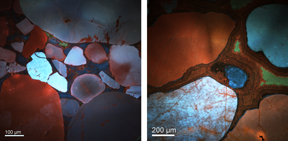
Reveal overgrowth and cementation processes
Color cathodoluminescence images reveal cementation processes in reservoir rocks. In the left image you can see low temperature quartz cement (bluish) and a later (hotter) overprint (reddish) along grain boundaries in the overgrowth. In the right image, you can see well developed overgrowth cement with multiple zoning. Image courtesy of Dr. Juergen Schieber, Indiana University Shale. Acquired on the ChromoCL2™ system.
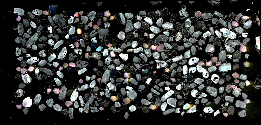
Measure trace elements, internal structures, and zoning
Here you can see a large collection of zircon grains were imaged to reveal full zonation structure and late rims that are only observable with cathodoluminescence. Field stitching software was used to create a 70 million pixel image with a field of view that is several millimeters in size. Results demonstrate that exact pixel registration between all signals allows you to directly correlate between topography, composition and trace element chemistry over large areas. Sample courtesy of Geological Survey Finland.
Analyze microfractures and fluid flow
Distribution of microfractures in geological materials can provide insight into emplacement events, such as damage associated with tectonic faulting. The below image shows how cathodoluminescence can be a valuable tool to reveal microfractures under high spatial resolution conditions (low accelerating voltage, low CL signal). Image courtesy of After Laubach et al., Journal of Structural Geology 2005.
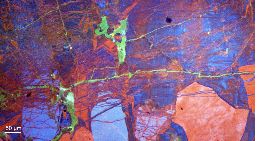
Cathodoluminescence provides unrivaled sensitivity to trace element composition so you can observe chemical overprinting where fluid flow along cracks has caused alteration in a polycrystalline diamond sample.
Identify minerals, phases, and their distribution
These results demonstrate the cathodoluminescence can reveal mineral attributes that are not available through alternative techniques. When compared, an EDS/BSE analysis (left) shows uniform distribution of quartz across the sample, while the secondary electron image (right) shows that there are a few large grains of authigenic and detrial quartz.

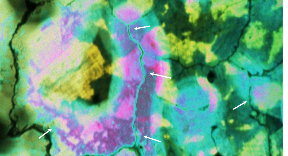
Reconstruct geological processes
To better understand the genesis of polycrystalline diamonds, you can compare the cathodoluminescence spectral characteristics to determine the cause of irradiation or point defects. For this sample, (a) a composite cathodoluminescence image of polycrystalline diamond was created using red, green, and blue spectrally filtered cathodoluminescence images. When cross-correlated with spectroscopy results, individual elements or substances were identified within the sample. Results showed a radioactive fluid ingress along grain boundaries lead to the radiation halo effects that were observed (yellow). Data courtesy of Dr. E. Vicenzi, Smithsonian Institute.
 at 50 spectra/s. The spectrum image gathered around the radiation halo allows the defects to be identified.")